. 2011 Aug 19;30(20):4157-70.
doi: 10.1038/emboj.2011.304.

Stabilizing the VE-cadherin-catenin complex blocks leukocyte extravasation and vascular permeability

Affiliations

Stabilizing the VE-cadherin-catenin complex blocks leukocyte extravasation and vascular permeability

Dörte Schulte et al. EMBO J. .

Abstract

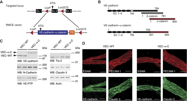
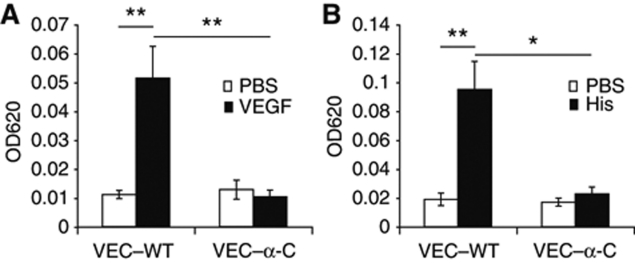
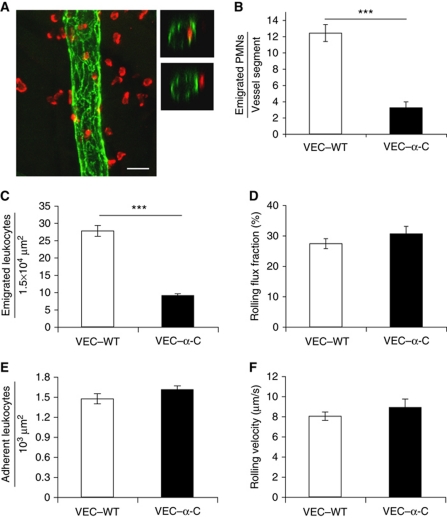
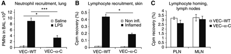
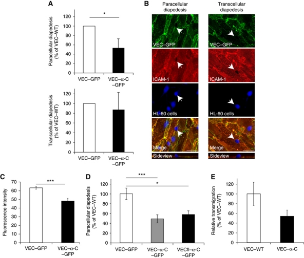
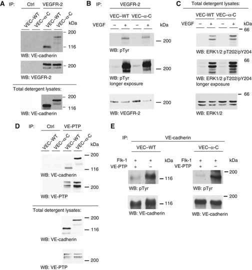
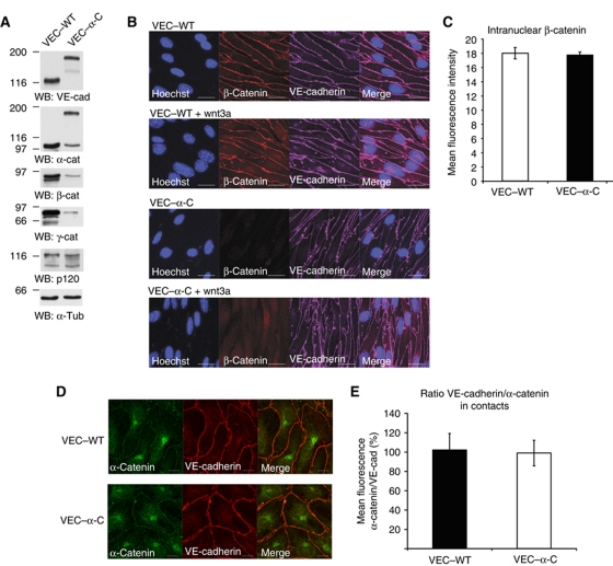
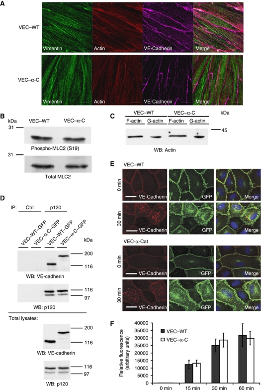
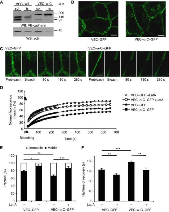
To determine whether leukocytes need to open endothelial cell contacts during extravasation, we decided to generate mice with strongly stabilized endothelial junctions. To this end, we replaced VE-cadherin genetically by a VE-cadherin-α-catenin fusion construct. Such mice were completely resistant to the induction of vascular leaks by VEGF or histamine. Neutrophil or lymphocyte recruitment into inflamed cremaster, lung and skin were strongly inhibited in these mice, documenting the importance of the junctional route in vivo. Surprisingly, lymphocyte homing into lymph nodes was not inhibited. VE-cadherin-α-catenin associated more intensely with the actin cytoskeleton as demonstrated by its membrane mobility and detergent extractability. Our results establish the junctional route as the main pathway for extravasating leukocytes in several, although not in all tissues. Furthermore, in these tissues, plasticity of the VE-cadherin-catenin complex is central for the leukocyte diapedesis mechanism.


Conflict of interest statement

The authors declare that they have no conflict of interest.

Figures

Figure 1
Figure 1
Generation of VE-cadherin–α-catenin knock-in mice. (A) Schematic illustration of targeting exon 2 (Ex2) of the VE-cadherin locus by recombinase-mediated cassette exchange (RMCE). Two incompatible LoxP sites (LoxP and Lox2272) were positioned to flank exon 2 in order to replace this exon in a second step with a replacement cassette flanked with the same Lox sites and containing VEC–α-C cDNA followed by a polyA-transcriptional stop (TS) cassette. (B) Schematic illustration of VE-cadherin, β-catenin, α-catenin and the VEC–α-C fusion protein depicting the extracellular (EC 1–5) and transmembrane (TM) domains of VE-cadherin. Numbers refer to amino-acid positions. (C) Western blot (WB) of SDS-urea extracts of lung tissue of homozygous adult VEC–WT and VEC–α-C mice, immunoblotted for VE-cadherin, N-cadherin, VE-PTP, Tie-2, claudin 5 and actin. Molecular weight markers are indicated on the right. Results of two mice for each genotype are shown. (D) Whole-mount stainings of cremaster muscles from VEC–WT and VEC–α-C mice using antibodies against VE-cadherin, ESAM, PECAM-1 and claudin 5. Projections of z-stacks taken on an LSM 510 Zeiss Meta, bars=20 μm. Figure source data can be found with the Supplementary Information.
Figure 2
Figure 2
VE-cadherin–α-catenin blocks VEGF and histamine-induced permeability in vivo. VEC–WT and VEC–α-C mice were intravenously injected with Evans Blue and 10 min later intradermally with PBS for controls (open bars) and either VEGF (A) or histamine (B) (black bars). Thirty minutes later, skin areas were excised and the dye was extracted and quantified. One representative experiment out of three independent experiments with at least four mice per group is shown. *P⩽0.05; **P⩽0.01; (A) t-test; (B) Mann rank Sum Test.
Figure 3
Figure 3
Extravasation of leukocytes into IL-1β-stimulated cremaster muscle is strongly reduced in VE-cadherin–α-catenin mice. (A, B) Confocal fluorescence microscopy was used to determine the number of extravasated neutrophils 3 h after intrascrotal IL-1β injection. (A) Representative images of whole mount stainings of cremaster labelled for PECAM-1 (green) and MRP-14 (red) to visualize endothelial cell contacts and neutrophils, respectively. Left panel: longitudinal vessel segment typically used for the 3D analysis and evaluation. Right panels: optical cross-sections of a venule depicting a neutrophil inside (upper right panel) and outside (lower right panel) the vessel. Projections of z-stacks taken on an LSM Zeiss 510 Meta, bar=20 μm. (B) Numbers of extravasated neutrophils in VEC–WT and VEC–α-C mice located within 50 μm next to the vessel were counted and are given per vessel segment (length 150 μm). A total of 60 (VEC–WT) and 66 (VEC–α-C) vessel segments from three mice of each group were analysed. (CF) Intravital microscopy of neutrophil extravasation in venules of the cremaster muscle 4 h after intrascrotal administration of IL-1β. (C) Numbers of extravasated leukocytes in cremaster venules of VEC–WT and VEC–α-C mice per 1.5 × 104 μm2 tissue area determined by reflected light oblique transillumination microscopy. (D) Rolling flux fraction, (E) number of adhering leukocytes per 103 μm2 of venule surface area, (F) rolling velocity. A total of 28 or 35 vessels from 4 VEC–WT and 5 VEC–α-C mice were analysed. Haemodynamic parameters are given in Supplementary Table S6, showing that peripheral neutrophil counts, enhanced under inflammatory conditions, were similar in VEC–WT and VEC–α-C mice. ***P⩽0.001; Mann Rank Sum Test.
Figure 4
Figure 4
VE-cadherin–α-catenin strongly reduces extravasation of neutrophils or lymphocytes into inflamed lung or skin, but not lymphocyte homing. (A) LPS induced PMN recruitment into inflamed lungs. The number of neutrophils in the bronchoalveolar lavage (BAL) of VEC–WT and VEC–α-C mice was analysed using flow cytometry 4 h after inhalation of LPS (black bars) or NaCl (open bars). Four mice were analysed per group. (B) DTH response in the skin was analysed by injection of radiolabelled, in vivo-activated lymphocytes from VEC–WT mice into DNFB-sensitized VEC–WT and VEC–α-C mice. Immigration of cells into the non-inflamed control ears (open bars) and inflamed ears (black bars) was analysed 5 h after lymphocyte injection. The depicted experiment was performed with six VEC–WT and four VEC–α-C mice and is representative of two separate experiments. (C) Radiolabelled naive lymphocytes from VEC–WT mice were injected intravenously into VEC–WT and VEC–α-C mice and the percentage of the total injected radioactivity in peripheral lymph nodes (PLN) and mesenteric lymph nodes (MLN) was determined 3 h later; n=5 (VEC–WT) and n=6 (VEC–α-C). One representative experiment out of two independent experiments is shown. *P⩽0.05; ***P⩽0.001, t-test.
Figure 5
Figure 5
VE-cadherin–α-catenin strongly reduces paracellular, but not transcellular diapedesis. (A) VEC–GFP or VEC–α-C–GFP was expressed by adenoviral vectors in HUVEC and paracellular and transcellular diapedesis were analysed as described in Materials and methods. DMSO differentiated granulocytic-HL60 cells were incubated with adenovirus transduced, TNF-α activated HUVEC for 20 min followed by fixation, staining and scoring of paracellular (top) and transcellular (bottom) diapedesis. In each case, the number of neutrophils migrating across VEC–GFP infected cells was set to 100%. A total of 39 or 44 areas containing 1090 or 1042 HL60 cells (including all loosely attached, adhering, transmigrating and transmigrated cells) were analysed for VEC–GFP and VEC–α-C–GFP expressing HUVEC, respectively. In all, 14% of these HL60 cells were transmigrating through VEC–GFP expressing HUVEC of which 96% migrated paracellular and 4% transcellular. (B) Representative immunofluorescence images of granulocytic-HL60 cells migrating through the transcellular or the paracellular route, endothelial cell contacts stained for VEC–GFP (green), HL60 stained by Cell Tracker (blue), apical endothelial cell surface stained for ICAM-1 (red). Projections of z-stacks taken on an LSM Zeiss 510 Meta, bars=20 μm. (C) Quantification of the expression levels of VEC–GFP and VEC–α-C–GFP by determining fluorescence intensity in 75 randomly selected ROIs of 50 μm2 at sites of cell contacts in HUVEC used for transmigration assays. (D) Transduced, TNF-α activated HUVEC cells expressing VEC–GFP, VEC–α-C–GFP or VECfl–α-C–GFP were incubated with granulocytic-HL60 cells for 20 min followed by fixation and staining. Paracellular migration through VEC–GFP expressing cells was set to 100%. A total of 18, 20 and 11 stacks containing 421, 426 and 302 HL60 cells with 74, 40 and 26 paracellular migrating cells were counted for VEC–GFP, VEC–α-C–GFP and VECfl–α-C–GFP expressing HUVEC, respectively. (E) Mouse PMNs were allowed to transmigrate for 30 min through TNF-α-stimulated endothelioma cells from VEC–WT and VEC–α-C mice grown on 3 μm pore size transwell filters. Transmigration through VEC–WT cells was set to 100%. *P⩽0.05, ***P⩽0.001; (A, D) Mann Rank Sum Test, (C, E) t-test.
Figure 6
Figure 6
VEGFR-2 signalling and VE-cadherin/VE-PTP interactions are not affected by fusion of VE-cadherin with α-catenin. (A) Cell lysates of confluent endothelioma cells generated from VEC–WT or VEC–α-C mice were immunoprecipitated (IP) with control antibodies (Ctrl) or antibodies against VEGFR-2 and precipitates were analysed by western blotting (WB) with antibodies to VE-cadherin and VEGFR-2, documenting association of VEGFR-2 with VE-cadherin and VEC–α-C. Note: Total detergent lysates contained less solubilized VEC–α-C than VE-cadherin protein (see below). (B, C) Confluent VEC–WT and VEC–α-C endothelioma cells were stimulated with VEGF (100 ng/ml) for 5 min, followed by (B) immunoblotting of immunoprecipitated VEGFR-2 with antibodies against phosphotyrosine (pTyr) and VEGFR-2; and (C) against phospho-Erk1/2 (pT202/pY204) and Erk1/2. Molecular weight markers are indicated on the right. (D) Anti-VE-PTP and control (Ctrl) immunoprecipitates from VEC–WT and VEC–α-C endothelioma cells (top panel) or total cell lysates (bottom panel) were immunoblotted for VE-cadherin or for VE-PTP (as indicated). (E) COS cells were transfected with VEC–WT (left) or VEC–α-C (right) and co-transfected with Flk1 and with (+) or without (−) VE-PTP. VE-cadherin immunoprecipitates were immunoblotted for phosphotyrosine (pTyr) or for VE-cadherin. Figure source data can be found with the Supplementary Information.
Figure 7
Figure 7
Catenin expression and subcellular distribution in VEC–WT and VEC–α-C cells. (A) Triton lysates of endothelioma cells established from VEC–WT and VEC–α-C mice were immunoblotted for VE-cadherin, α-catenin, β-cat, plakoglobin, p120 and α-tubulin (as indicated). (B) VEC–WT and VEC–α-C endothelioma cells were treated for 14 h with or without wnt3a, fixed, permeabilized and stained for nuclei, VE-cadherin and β-catenin. (C) Quantification of nuclear β-catenin staining after wnt3a stimulation, t-test. (D) Immunofluorescence staining of VEC–WT and VEC–α-C endothelioma cells for VE-cadherin and α-catenin. (E) Quantification of the ratio of VE-cadherin/α-catenin staining at cell contact as shown in (D), t-test. Figure source data can be found with the Supplementary Information.
Figure 8
Figure 8
Cytoskeletal organization and VE-cadherin endocytosis are not affected by VEC–α-C. (A) Immunofluorescence staining of VEC–WT and VEC–α-C endothelioma cells for vimentin, VE-cadherin and actin (as indicated). (B) Western blots of cell lysates of the same cells for total myosin light chain (MLC2) and its phosphorylated form. (C) F-actin and G-actin were isolated from the same cells and detected by immunoblotting. (D) Lung lysates of VEC–WT and VEC–α-C mice were immunoprecipitated for p120 or with control antibodies (Ctrl) and precipitates were immunoblotted for p120 or VE-cadherin. Bottom: respective immunoblots of total lung lysate aliquots. (E, F) Endocytosis of VE-cadherin. (E) VE-cadherin–GFP (VEC–WT) or VE-cadherin–α-catenin–GFP (VEC–α-C) was expressed by adenoviral vectors in HUVEC cells, incubated with an anti-VE-cadherin antibody at 4°C, and after washing away excess of antibody, cells were allowed to endocytose VE-cadherin for 0 or 30 min. Cells were fixed, permeabilized and stained with a secondary antibody against the first antibody (red). GFP is seen in green, merge to the right. (F) Quantification of intracellular (red) VE-cadherin staining of HUVEC that had endocytosed VE-cadherin–GFP (VEC–WT) or VE-cadherin–α-catenin–GFP (VEC–α-C) for 0, 15, 30 or 60 min, t-test. Figure source data can be found with the Supplementary Information.
Figure 9
Figure 9
Fusion of VE-cadherin with α-catenin decreases detergent extractability and membrane mobility. (A) Embryonic endothelioma cell lines established from mutant VEC–WT and VEC–α-C embryos were extracted with 1% NP-40 followed by immunoblot analysis of soluble (sol) and insoluble (is) fractions as described in Materials and methods. Molecular weight markers are indicated on the right. (B) Representative images of HUVEC expressing VEC–GFP or VEC–α-C–GFP used for FRAP analysis. Note the straightened cell contacts in cells expressing VEC–α-C–GFP compared with VEC–GFP. Bars=10 μm. (C) Representative images of cell contact areas of HUVEC expressing VEC–GFP or VEC–α-C–GFP at indicated time points prior and post photobleaching. Bars=5 μm. (D) Time course of fluorescence recovery at bleached areas of cell contacts of vehicle-treated HUVEC expressing VEC–GFP (black triangles, 24 areas analysed) or VEC–α-C–GFP (black boxes, 32 areas analysed) and the respective latrunculin A treated, transduced HUVEC (+LatA, open triangles or boxes, 19 and 14 areas analysed). (E, F) Mobile (white) and immobile (black) fraction (E) and recovery halftime of VEC–GFP or VEC–α-C–GFP (F) in transduced HUVEC treated with vehicle or latrunculin A (as indicated) calculated from the curves shown in (D). *P⩽0.05; **P⩽0.01; ***P⩽0.001, t-test. Figure source data can be found with the Supplementary Information.

Similar articles

Cited by

References

    1. Abe K, Takeichi M (2008) EPLIN mediates linkage of the cadherin catenin complex to F-actin and stabilizes the circumferential actin belt. Proc Natl Acad Sci USA 105: 13–19 - PMC - PubMed
    1. Alksne JF (1959) The passage of colloidal particles across the dermal capillary wall under the influence of histamine. Q J Exp Physiol Cogn Med Sci 44: 51–66 - PubMed
    1. Allingham MJ, van Buul JD, Burridge K (2007) ICAM-1-mediated, Src- and Pyk2-dependent vascular endothelial cadherin tyrosine phosphorylation is required for leukocyte transendothelial migration. J Immunol 179: 4053–4064 - PubMed
    1. Anderson AO, Anderson ND (1976) Lymphocyte emigration from high endothelial venules in rat lymph nodes. Immunology 31: 731–748 - PMC - PubMed
    1. Baluk P, Bolton P, Hirata A, Thurston G, McDonald DM (1998) Endothelial gaps and adherent leukocytes in allergen-induced early- and late-phase plasma leakage in rat airways. Am J Pathol 152: 1463–1476 - PMC - PubMed

Publication types

MeSH terms