. 2011 Aug 21;477(7362):111-4.
doi: 10.1038/nature10349.

Solution structure of a minor and transiently formed state of a T4 lysozyme mutant

Affiliations

Solution structure of a minor and transiently formed state of a T4 lysozyme mutant

Guillaume Bouvignies et al. Nature. .

Abstract

Proteins are inherently plastic molecules, whose function often critically depends on excursions between different molecular conformations (conformers). However, a rigorous understanding of the relation between a protein's structure, dynamics and function remains elusive. This is because many of the conformers on its energy landscape are only transiently formed and marginally populated (less than a few per cent of the total number of molecules), so that they cannot be individually characterized by most biophysical tools. Here we study a lysozyme mutant from phage T4 that binds hydrophobic molecules and populates an excited state transiently (about 1 ms) to about 3% at 25 °C (ref. 5). We show that such binding occurs only via the ground state, and present the atomic-level model of the 'invisible', excited state obtained using a combined strategy of relaxation-dispersion NMR (ref. 6) and CS-Rosetta model building that rationalizes this observation. The model was tested using structure-based design calculations identifying point mutants predicted to stabilize the excited state relative to the ground state. In this way a pair of mutations were introduced, inverting the relative populations of the ground and excited states and altering function. Our results suggest a mechanism for the evolution of a protein's function by changing the delicate balance between the states on its energy landscape. More generally, they show that our approach can generate and validate models of excited protein states.


Figures

Figure 1
Figure 1. L99A T4L exchanges between ground (visible) and excited (invisible) states, each with distinct conformations.
a, Plot of ΔϖRMS=1Ni(ΔϖiΔϖi,STD)2 as a function of residue, where Δω̄i is the shift difference in p.p.m. between states, Δω̄i,STD is a nucleus specific value that corresponds to the range of shift values (1 s.d.) that are observed in a database of protein chemical shifts (http://www.bmrb.wisc.edu) for the nucleus in question (1HN, 15N, 13Cα, 1Hα and 13C′) and N is the number of nuclei ≤5 that are included in the average. Significant Δω̄RMS differences are localized to a pair of regions (100–120, 132–146) that are highlighted in grey. The secondary structure of the ground state of L99A T4L is illustrated. b, Values of Δω̄RMS colour-coded onto the X-ray structure of L99A T4L (PDB: 3DMV), ranging from blue (Δω̄RMS = 0) to red (Δω̄RMS > 0.7). The mesh surface indicates the position of the cavity formed by the Leu to Ala substitution at position 99. c, S2 values forthe backbone amide groupsin the ground (blue) and excited(red)states of L99A T4L as predicted by the RCI approach. d, Helix propensity values, predicted using TALOS+ (ref. 29), highlighting important changes in secondary structure between ground (blue) and excited (red) L99A T4L conformers.
Figure 2
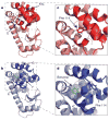
Figure 2. The structure of the invisible, excited state of L99A T4L
a, Superposition of the 10 lowest energy structures of the L99A T4L excited state. The locations of helices E, F and G are indicated, along with the side chain of Phe 114 (see c). Only residues 100–120 and 132–146 were allowed to deviate from the L99A T4L ground state X-ray structure in calculations of the excited conformer (Methods). b, Ground state structure of L99A T4L, showing helices E, F and G and the position of Phe 114 (PDB: 3DMV) or of benzene (green; PDB: 3DMX) when it is bound inside the cavity (see d). c, d, Expanded regions of the excited state (c) and ground state (d) structures, focusing on the differences between helices F and G and the position of Phe 114.
Figure 3
Figure 3. Hydrophobic ligands do not bind the excited state of L99A T4L
a, Selected region of the 1H–15N correlation map from a magnetization exchange experiment recorded on L99A, G113A T4L at 1 °C, showing separate peaks for the ground (G) and excited (E) states. A pair of data sets are obtained, with the mixing time (TMIX = 50 ms) recorded before or after the 15N chemical shift evolution period, and the data sets subtracted so that diagonal- (cross-) peaks are positive (negative). b, Correlation between Δω̄N values measured directly from the spectrum in a (y axis) and corresponding values from CPMG relaxation dispersion measurements of L99A T4L (25 °C; x axis). c, Magnetization exchange spectrum (TMIX = 40 ms) recorded on an L99A, G113A T4L sample with a 1:1 molar equivalent of benzene, focusing on Met 102 that shows well resolved correlations from ground, excited and benzene-bound (B) states. d, Intensity of auto- and cross-peaks for residue Met 102 from magnetization exchange experiments recorded as a function of TMIX (red circles), along with the best fit of the data (solid lines) to the exchange model of e. Values of rates of exchange, kij, are indicated.
Figure 4
Figure 4. The delicate balance between states on the energy landscape can be readily manipulated through mutation, providing a path for protein evolvability
a–c, Selected regions from 1H–13C HSQC spectra (recorded at 1 °C) of L99A T4L (a), L99A, G113A T4L (b) and L99A, G113A, R119P T4L (c), with the peaks from the ground and excited states coloured in blue and red, respectively. d–f, Corresponding energy landscapes, showing the structures of the ground and excited states and their fractional populations.

References

    1. Karplus M, Kuriyan J. Molecular dynamics and protein function. Proc Natl Acad Sci USA. 2005;102:6679–6685. - PMC - PubMed
    1. Boehr DD, McElheny D, Dyson HJ, Wright PE. The dynamic energy landscape of dihydrofolate reductase catalysis. Science. 2006;313:1638–1642. - PubMed
    1. Henzler-Wildman KA, et al. Intrinsic motions along an enzymatic reaction trajectory. Nature. 2007;450:838–844. - PubMed
    1. Eriksson AE, Baase WA, Wozniak JA, Matthews BW. A cavity-containing mutant of T4 lysozyme is stabilized by buried benzene. Nature. 1992;355:371–373. - PubMed
    1. Mulder FAA, Mittermaier A, Hon B, Dahlquist FW, Kay LE. Studying excited states of protein by NMR spectroscopy. Nature Struct Biol. 2001;8:932–935. - PubMed

Publication types