. 2011 Jul 1;144(2):379-404.
doi: 10.1007/s10955-011-0214-y.

Statistical Mechanics of Nucleosomes Constrained by Higher-Order Chromatin Structure

Affiliations

Statistical Mechanics of Nucleosomes Constrained by Higher-Order Chromatin Structure

Răzvan V Chereji et al. J Stat Phys. .

Abstract

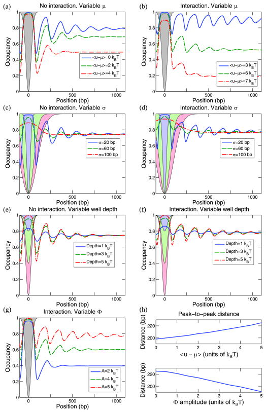
Eukaryotic DNA is packaged into chromatin: one-dimensional arrays of nucleosomes separated by stretches of linker DNA are folded into 30-nm chromatin fibers which in turn form higher-order structures (Felsenfeld and Groudine in Nature 421:448, 2003). Each nucleosome, the fundamental unit of chromatin, has 147 base pairs (bp) of DNA wrapped around a histone octamer (Richmond and Davey in Nature 423:145, 2003). In order to describe how chromatin fiber formation affects nucleosome positioning and energetics, we have developed a thermodynamic model of finite-size particles with effective nearest-neighbor interactions and arbitrary DNA-binding energies. We show that both one-and two-body interactions can be extracted from one-particle density profiles based on high-throughput maps of in vitro or in vivo nucleosome positions. Although a simpler approach that neglects two-body interactions (even if they are in fact present in the system) can be used to predict sequence determinants of nucleosome positions, the full theory is required to disentangle one- and two-body effects. Finally, we construct a minimal model in which nucleosomes are positioned primarily by steric exclusion and two-body interactions rather than intrinsic histone-DNA sequence preferences. The model reproduces nucleosome occupancy patterns observed over transcribed regions in living cells.


Figures

Fig. 1
Fig. 1
(Color online) A typical nucleosome configuration near the transcription start site (TSS). The blue background represents in vivo nucleosome occupancy from Zawadzki et al. [18], averaged over all genes. The occupancy profile is normalized by its average in the [−500, 500] bp window around the TSS. The intensity of the blue color is proportional to the degree of nucleosome localization
Fig. 2
Fig. 2
(Color online) A typical configuration of 6 nucleosomes. In this toy representation, each nucleosome covers 4 bps of DNA sequence (represented by colored boxes) which gives the one-body energy of the nucleosome, u. The one-body energy is represented by gray bars. For simplicity, the one-body energy (shown as the height of the gray bar) is assumed to be entirely determined by the base pair located at the starting position of the nucleosome. In more realistic scenarios the one-body energy is a function of the entire sequence occupied by the nucleosome. The two-body interaction Φ (i, j) acts only between neighboring nucleosomes, with two indices i and j representing their starting positions
Fig. 3
Fig. 3
(Color online) (a) g(i, j) = n2(i, j)/n(i)n(j) is plotted for a representative subset of all initial positions i in a 104 bp DNA segment. The one-body energies are randomly sampled from a Gaussian distribution with a mean of 2.5 kB T and a standard deviation of 0.2 kB T, and 9 potential wells of depth 5 kB T are added at 1, 2, …, 9 ×10³ bp to model a strongly inhomogeneous system. n2(i, j) and n(i) are computed from one-and two-body energies using (3) and (4). (b) Plinker(Δ), obtained by averaging g(i, j) over all initial positions i. Note that Δ = j − (i + 147) represents the linker length between the two nucleosomes with starting positions i and j, respectively. (c) Exact (solid blue line) and predicted (dotted black line) two-body interactions. The predicted interaction was computed from the – ln(Plinker) curve (dashed green line) using (17)
Fig. 4
Fig. 4
(Color online) (a) One-body energies for a 10000 bp DNA segment. Sequence-dependent energy given by the 21-parameter model (26) (blue solid line), total energy given by the sum of the sequence-specific energies and 5 potential wells with 3 kBT depth at 1, 3, 5, 7, and 9 ×10³ bp designed to mimic the in vivo effects (green dash-dot-dot line). Energy predicted with a model that neglects two-body interactions (10) (red dashed line), energy predicted by fitting the 21-parameter model to the energies from (10) (light blue dash-dot line), a numerical solution of the full model which takes Φ into account (maroon dotted line). Inset: zoom-in on the region with one of the potential wells. (b) Exact two-body interaction Φ (blue solid line) and predicted interaction (17) (green dashed line). (c) Nucleosome occupancies. Occupancy generated by the exact sequence-specific one-body energy and the exact interaction (blue solid line), occupancy corresponding to the combined exact one-body energy (sequence-specific component and potential wells) and the exact interaction (green dash-dot-dot line). Predicted occupancy generated by the one-body energy from (10) and predicted Φ (red dashed line), occupancy generated using predicted sequence-dependent one-body energy (26) and predicted Φ (light blue dash-dot line), occupancy predicted using numerically computed one-body energies from the full model and predicted Φ (maroon dotted line). One-body energies and nucleosome occupancies are shown in a [600,1100] bp window
Fig. 5
Fig. 5
(Color online) Symmetric Gaussian barrier. Occupancy profiles for the following scenarios: variable chemical potential μ (a) and (b), variable barrier width (c) and (d), and variable barrier height (e) and (f). Unless otherwise specified in the legend, the barrier heights are 5 kBT, σ = 30 bp, and 〈uμ〉 = 5 kBT [in panel (c) 〈uμ〉 = 1 kBT]. Panels (b), (d) and (f) have a two-body interaction Φ(Δ) = A cos (2π Δ/10) exp(−Δ/50), with A = 5 kBT. (g) Occupancy profiles for variable interaction strength A. (h) Variation of the distance between +1 and +2 nucleosomes located immediately to the right of the barrier (computed as the distance between the maxima of their occupancy) as μ or A is varied. Upper panel: Φ = 0, lower panel: 〈u〉 − μ = 5 kBT
Fig. 6
Fig. 6
(Color online) Symmetric Gaussian well. Occupancy profiles for the scenarios described in Fig. 5. All the parameters not explicitly given in the legends are from Fig. 5. In particular, well depths have the same magnitude as the heights of the corresponding barriers
Fig. 7
Fig. 7
(Color online) Asymmetric Gaussian barrier. Occupancy profiles for the scenarios described in Fig. 5. Unless otherwise specified in the legend, the barrier heights are 5 kBT, σL = 70 bp, σR = 30 bp, and 〈uμ〉 = 5 kBT [in panel (c) 〈uμ〉 = 1 kBT]
Fig. 8
Fig. 8
(Color online) Average nucleosome occupancy in the vicinity of transcription start and termination sites (TSS and TTS, respectively). Each occupancy profile is normalized by its average in the [−500, 500] bp window. (a), (b): Nucleosome occupancy observed in vivo (YPD medium) and in vitro by Kaplan et al. [12] and in vitro by Zhang et al. [13], and predicted using a 21-parameter N = 2 position-independent model, a minimal model in which nucleosomes are localized purely by means of sequence-independent potential barriers (Fig. 9), and a combined model in which sequence-specific energies from the 21-parameter N = 2 model are added to the barriers from Fig. 9. The two-body potential is turned off. Note that in Ref. [13] DNA was mixed with histones in a 1:1 mass ratio which is close to the in vivo value, while in Ref. [12] the ratio was 0.4:1, resulting in deeper NDRs. (c), (d): Nucleosome occupancy observed in vivo by Zawadzki et al. [18] and Mavrich et al. [16], and predicted using the 21-parameter N = 2 position-independent model, the minimal model, and the combined model. The two-body potential is given by Φ(Δ) = A cos (2π Δ/10) exp(−Δ/50), with A = 5 kBT
Fig. 9
Fig. 9
(Color online) The one-body energy profiles used in Figs. 8 and 10. The 5′ asymmetric barrier has σleft = 80 bp and σright = 30 bp. The 3′ symmetric barrier has σ = 80 bp. Solid blue line: barriers used in the in vivo minimal model without two-body interactions [Figs. 8(a) and 8(b), Fig. 10]. Dash-dot green line: barriers used in the in vivo combined model without two-body interactions [Figs. 8(a) and 8(b), Fig. 10]. Dashed yellow line: barriers used in the in vivo minimal model with two-body interactions [Figs. 8(c) and 8(d)]. Dotted light blue line: barriers used in the in vivo combined model with two-body interactions [Figs. 8(c) and 8(d)]. The energy landscapes shown in the figure are shifted vertically so that 〈uμ〉 = 0.56 kBT in the minimal model without two-body interactions, 0.62 kBT in the combined model without two-body interactions, 4.49 kBT in the minimal model with two-body interactions, and 4.62 kBT in the combined model with two-body interactions
Fig. 10
Fig. 10
(Color online) Heat maps of nucleosome occupancy around TSS and TTS for 5747 S. cerevisiae genes. In vivo nucleosomes (YPD medium) [12] (a) and (b), N = 2 position-independent model (c) and (d), minimal model (e) and (f), combined model (g) and (h). The minimal model is constructed by placing potential barriers from Fig. 9 at the end of each gene onto an otherwise flat one-body energy landscape without two-body interactions. The combined model is constructed by adding sequence-specific energies from the 21-parameter N = 2 position-independent model (which have standard deviation of 0.61 kBT genome-wide) to the minimal model. The occupancy for each gene is normalized by the average occupancy in the [−500, 500] bp window. The experimental data [(a) and (b)] are smoothed with a 2D Gaussian kernel (σX = 1 bp and σY = 2 genes). The genes are sorted in each panel in the order of increasing variance of the occupancy. The genome-wide average occupancies are 0.15 [(a) and (b)], 0.20 [(c) and (d)], 0.75 [(e) and (f)], and 0.72 [(g) and (h)]

References

    1. Felsenfeld G, Groudine M. Nature. 2003;421:448. - PubMed
    1. Richmond TJ, Davey CA. Nature. 2003;423:145. - PubMed
    1. Struhl K. Cell. 1999;98:1. - PubMed
    1. Mellor J. Trends Genet. 2006;22:320. - PubMed
    1. Li B, Carey M, Workman JL. Cell. 2007;128:707. - PubMed

LinkOut - more resources