

Integrated proteomic and transcriptomic investigation of the acetaminophen toxicity in liver microfluidic biochip
- PMID: 21857903
- PMCID: PMC3152546
- DOI: 10.1371/journal.pone.0021268
Integrated proteomic and transcriptomic investigation of the acetaminophen toxicity in liver microfluidic biochip
Abstract
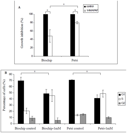
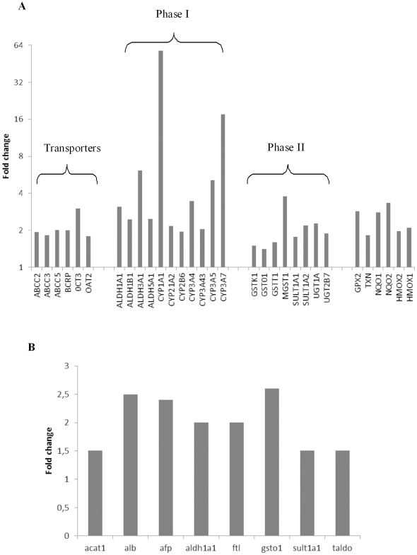
Microfluidic bioartificial organs allow the reproduction of in vivo-like properties such as cell culture in a 3D dynamical micro environment. In this work, we established a method and a protocol for performing a toxicogenomic analysis of HepG2/C3A cultivated in a microfluidic biochip. Transcriptomic and proteomic analyses have shown the induction of the NRF2 pathway and the related drug metabolism pathways when the HepG2/C3A cells were cultivated in the biochip. The induction of those pathways in the biochip enhanced the metabolism of the N-acetyl-p-aminophenol drug (acetaminophen-APAP) when compared to Petri cultures. Thus, we observed 50% growth inhibition of cell proliferation at 1 mM in the biochip, which appeared similar to human plasmatic toxic concentrations reported at 2 mM. The metabolic signature of APAP toxicity in the biochip showed similar biomarkers as those reported in vivo, such as the calcium homeostasis, lipid metabolism and reorganization of the cytoskeleton, at the transcriptome and proteome levels (which was not the case in Petri dishes). These results demonstrate a specific molecular signature for acetaminophen at transcriptomic and proteomic levels closed to situations found in vivo. Interestingly, a common component of the signature of the APAP molecule was identified in Petri and biochip cultures via the perturbations of the DNA replication and cell cycle. These findings provide an important insight into the use of microfluidic biochips as new tools in biomarker research in pharmaceutical drug studies and predictive toxicity investigations.
Conflict of interest statement
Figures

Similar articles
-
Predictive toxicology using systemic biology and liver microfluidic "on chip" approaches: application to acetaminophen injury.Toxicol Appl Pharmacol. 2012 Mar 15;259(3):270-80. doi: 10.1016/j.taap.2011.12.017. Epub 2011 Dec 24. Toxicol Appl Pharmacol. 2012. PMID: 22230336
-
Investigation of acetaminophen toxicity in HepG2/C3a microscale cultures using a system biology model of glutathione depletion.Cell Biol Toxicol. 2015 Jun;31(3):173-85. doi: 10.1007/s10565-015-9302-0. Epub 2015 May 9. Cell Biol Toxicol. 2015. PMID: 25956491
-
Investigation of ifosfamide nephrotoxicity induced in a liver-kidney co-culture biochip.Biotechnol Bioeng. 2013 Feb;110(2):597-608. doi: 10.1002/bit.24707. Epub 2012 Aug 28. Biotechnol Bioeng. 2013. PMID: 22887128
-
Metabolic phenotyping applied to pre-clinical and clinical studies of acetaminophen metabolism and hepatotoxicity.Drug Metab Rev. 2015 Feb;47(1):29-44. doi: 10.3109/03602532.2014.982865. Epub 2014 Dec 23. Drug Metab Rev. 2015. PMID: 25533740 Review.
-
Transcriptomic studies on liver toxicity of acetaminophen.Drug Dev Res. 2014 Sep;75(6):419-23. doi: 10.1002/ddr.21227. Drug Dev Res. 2014. PMID: 25195586 Review.
Cited by
-
Human-induced pluripotent stem cell-derived cardiomyocytes, 3D cardiac structures, and heart-on-a-chip as tools for drug research.Pflugers Arch. 2021 Jul;473(7):1061-1085. doi: 10.1007/s00424-021-02536-z. Epub 2021 Feb 24. Pflugers Arch. 2021. PMID: 33629131 Free PMC article. Review.
-
Novel in vitro and mathematical models for the prediction of chemical toxicity.Toxicol Res (Camb). 2013 Jan 1;2(1):40-59. doi: 10.1039/c2tx20031g. Epub 2012 Sep 5. Toxicol Res (Camb). 2013. PMID: 26966512 Free PMC article. Review.
-
Translational biomarkers of acetaminophen-induced acute liver injury.Arch Toxicol. 2015 Sep;89(9):1497-522. doi: 10.1007/s00204-015-1519-4. Epub 2015 May 17. Arch Toxicol. 2015. PMID: 25983262 Free PMC article. Review.
-
Microfabricated Physiological Models for In Vitro Drug Screening Applications.Micromachines (Basel). 2016 Dec 15;7(12):233. doi: 10.3390/mi7120233. Micromachines (Basel). 2016. PMID: 30404405 Free PMC article. Review.
-
Blood gene expression profiling of an early acetaminophen response.Pharmacogenomics J. 2017 Jun;17(3):230-236. doi: 10.1038/tpj.2016.8. Epub 2016 Mar 1. Pharmacogenomics J. 2017. PMID: 26927286 Free PMC article. Clinical Trial.
References
-
- Ferrer-Dufol A, Menao-Guillen S. Toxicogenomics and clinical toxicology: An example of the connection between basic and applied sciences. Toxicol Lett. 2009;186:2–8. - PubMed
-
- Oberemm A, Ahr HJ, Bannasch P, Ellinger-Ziegelbauer H, Glückmann M, et al. Toxicogenomic analysis of N-nitrosomorpholine induced changes in rat liver: Comparison of genomic and proteomic responses and anchoring to histopathological parameters. Toxicol Appl Pharmacol. 2009;241:230–245. - PubMed
-
- Veenstra TD. Proteomic approaches in drug discovery. Drug Discovery Today: Technologies. 2006;3:433–440.
-
- Lee WM. Drug-Induced Hepatotoxicity. N Engl J Med. 2003;349:474–485. - PubMed
Publication types
MeSH terms
Substances
LinkOut - more resources
Full Text Sources
