. 2011;6(8):e21396.
doi: 10.1371/journal.pone.0021396. Epub 2011 Aug 3.

miR-34a regulates mouse neural stem cell differentiation

Affiliations

miR-34a regulates mouse neural stem cell differentiation

Márcia M Aranha et al. PLoS One. 2011.

Abstract

Background: MicroRNAs (miRNAs or miRs) participate in the regulation of several biological processes, including cell differentiation. Recently, miR-34a has been implicated in the differentiation of monocyte-derived dendritic cells, human erythroleukemia cells, and mouse embryonic stem cells. In addition, members of the miR-34 family have been identified as direct p53 targets. However, the function of miR-34a in the control of the differentiation program of specific neural cell types remains largely unknown. Here, we investigated the role of miR-34a in regulating mouse neural stem (NS) cell differentiation.

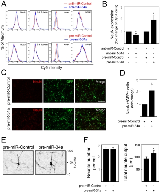
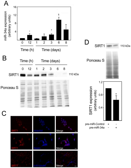
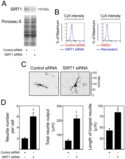
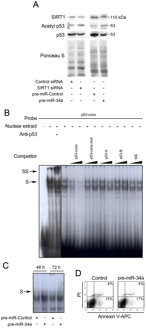
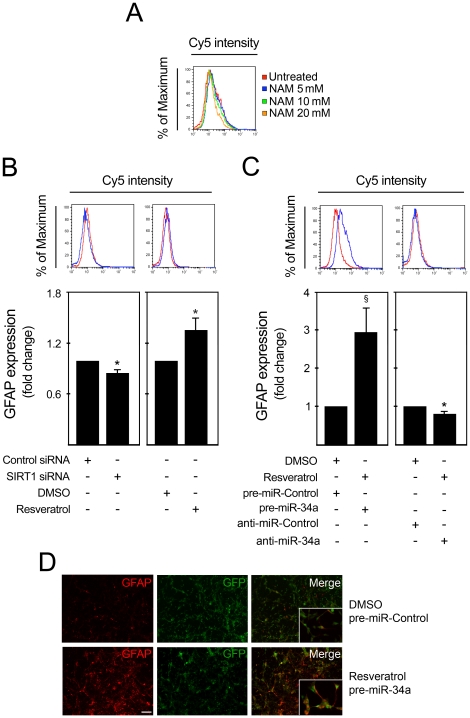
Methodology/principal findings: miR-34a overexpression increased postmitotic neurons and neurite elongation of mouse NS cells, whereas anti-miR-34a had the opposite effect. SIRT1 was identified as a target of miR-34a, which may mediate the effect of miR-34a on neurite elongation. In addition, acetylation of p53 (Lys 379) and p53-DNA binding activity were increased and cell death unchanged after miR-34a overexpression, thus reinforcing the role of p53 during neural differentiation. Interestingly, in conditions where SIRT1 was activated by pharmacologic treatment with resveratrol, miR-34a promoted astrocytic differentiation, through a SIRT1-independent mechanism.

Conclusions: Our results provide new insight into the molecular mechanisms by which miR-34a modulates neural differentiation, suggesting that miR-34a is required for proper neuronal differentiation, in part, by targeting SIRT1 and modulating p53 activity.


Conflict of interest statement

Competing Interests: The authors have declared that no competing interests exist.

Figures

Figure 1
Figure 1. miR-34a modulates the proportion of NeuN-positive cells and neurite outgrowth.
Mouse NS cells were transfected using 100 nM of either anti- or pre-miR-34a at 3 days, and collected after 24 and 72 h, respectively. Cells were subsequently labeled for Nestin, β-III Tubulin, NeuN and GFAP detection by flow cytometry. A. Representative histograms of Nestin, β-III Tubulin, NeuN and GFAP detection in anti-miR-control (red line) and anti-miR-34a-transfected cultures (blue line) (top), or in pre-miR-control (red line) and pre-miR-34a-transfected cultures (blue line) (bottom). In four independent experiments, similar alterations in the relative number of positive cells for each marker were observed. B. Quantification data of NeuN-positive cells assessed by flow cytometry after miR-34a modulation. C. Immunofluorescence detection of NeuN expression in 6 day cells transfected with pre-miR-Control and pre-miR-34a for 72 h. Scale bar, 50 µm. D. Quantification data of NeuN-positive cells assessed by immunocytochemistry after miR-34a modulation. E. Representative images of β-III Tubulin+ cells 48 h after pre-miR-transfection. Fluorescent images are shown in black and white and inverted for clarity. F. Neurite number and total neurite output are given to quantify the effect of miR-34a overexpression on cellular morphology. Neurites were manually traced using ImageJ v 1.43 and the NeuronJ plugin v 1.4.2. Results are shown as mean ± SEM. *p<0.05 from cells transfected with respective control.
Figure 2
Figure 2. SIRT1 expression levels decrease after miR-34a overexpression.
miR-34a expression was analyzed by quantitative Real Time-PCR using specific Taqman primers and GAPDH for normalization. Expression levels were calculated by the ΔΔCt method using undifferentiated cells as calibrator. SIRT1 expression in mouse NS cells was detected by immunoblotting at different times of differentiation or by immunocytochemistry at 2 days of differentiation. Cells transfected with either control or pre-miR-34a at 3 days of differentiation were also processed for SIRT1 detection by immunoblotting. A. Expression of miR-34a throughout the differentiation period. *p<0.05, p<0.01 and § p<0.001 compared to day 0 (undifferentiated cells). Data represent mean ± SEM of four independent experiments. B. Representative immunoblot showing a marked decrease in SIRT1 expression levels from day 3 of differentiation. Ponceau S was used as loading control. C. Immunofluorescence detection of cells labeled with anti-SIRT1, anti-Nestin and anti-β-III tubulin antibodies to visualize nuclear SIRT1 expression in neural progenitors and neuronal precursors, respectively. Hoechst 33258 staining was used to visualize cell nuclei. Scale bar, 10 µm. D. Representative immunoblot (top) and corresponding densitometry analysis (bottom) showing decreased SIRT1 expression in pre-miR-34a transfected cells. Data represent mean ± SEM of three independent experiments. *p<0.05 from cells transfected with control.
Figure 3
Figure 3. miR-34a may mediate neurite outgrowth by downregulating SIRT1 expression.
SIRT1 expression was modulated in mouse NS cells at 12 h after induction of differentiation. Inhibition of SIRT1 was achieved by transfecting cells with either scrambled control or 100 nM of SIRT1 siRNA, while activation of SIRT1 was performed by treating cells with either DMSO (control) or 5 µM of resveratrol. Cells were collected at 3 days, fixed and processed for NeuN labeling and detection by flow cytometry or for immunocytochemistry of β-III Tubulin and subsequent characterization of neurite elongation and branching. The blue line corresponds to SIRT1 modulation, while the red line corresponds to the respective control. A. Representative immunoblot showing decreased SIRT1 expression after transfection with SIRT1 siRNA. Ponceau S was used as loading control. B. SIRT1 modulation had no effect on the percentage of NeuN+ cells. C. Representative fluorescence microscopy image of β-III Tubulin immunocytochemistry after SIRT1 downregulation. Fluorescent images are shown in black and white and inverted for clarity. D. Neurite number, total neurite output and the length of longest neurite were determined to quantify the effect of SIRT1 downregulation on cellular morphology. Neurites were manually traced using ImageJ v 1.43 and the NeuronJ plugin v 1.4.2. Results are shown as mean ± SEM. § p<0.0001 from cells transfected with respective control.
Figure 4
Figure 4. miR-34a overexpression increases acetylation of p53 and p53-DNA binding activity.
Mouse NS cells at 3 days of differentiation were transfected with either SIRT1 siRNA or scrambled control for 48 h or with pre-miR-control or pre-miR-34a for 72 h. Cells were then collected for total and nuclear protein extraction or stained with Annexin-V-APC/PI to evaluate cell death. p53-overexpressing cells were used for supershift and competition experiments. Radio-labeled double-stranded oligonucleotide corresponding to the p53 consensus (p53 -cons) was used as a probe. A. Representative immunoblot showing increased p53 acetylation at lysine 379 after SIRT1 silencing or overexpression of miR-34a. Ponceau S was used as loading control. B. Representative EMSA showing the specificity of the complex formed with p53-cons probe. Supershift experiments were performed using an anti-p53 antibody (DO-1, Santa Cruz Biotechnology). Competition experiments were performed by adding 10- or 100-fold excess of unlabeled double-stranded oligonucleotides bearing a mutation in the consensus site (p53-cons-mut), or containing either two or one quarter-sites known to be consensus sites for p53 (p53-A and p53-B, respectively) or a non-specific sequence (NS). C. EMSA showing increased p53-DNA binding activity in pre-miR-34a transfected cells for 48 and 72 h. D. Representative Annexin V-APC/PI staining showing absence of cell death after miR-34a modulation.
Figure 5
Figure 5. miR-34a promotes astrocytic differentiation under resveratrol treatment.
SIRT1 expression was modulated in mouse NS cells after 12 h of induction of differentiation. Inhibition of SIRT1 was achieved by incubation with nicotinamide or by transfecting cells with either scrambled control or 100 nM of SIRT1 siRNA. SIRT1 was activated by treating cells with either DMSO (control) or 5 µM of resveratrol. Cells were collected at 3 days, fixed and processed for GFAP labeling and detection by flow cytometry and immunofluorescence. The blue line corresponds to SIRT1 modulation, while the red line represents the respective control. A. Incubation with nicotinamide, decreased the percentage of GFAP+ cells in a dose-dependent manner. B. Decreased percentage of GFAP+ cells after silencing of SIRT1 (left), and increased percentage of GFAP+ after SIRT1 activation (right). C. Under resveratrol treatment, overexpression of miR-34a resulted in increased proportion of GFAP+ cells (right), while miR-34a downregulation had the opposite effect (left). D. Immunofluorescence showing increased number of GFAP+ cells under resveratrol treatment and miR-34a overexpression (bottom) when compared with controls (top). Scale bar, 80 µm. Images taken with a magnification of 400× are shown in detail. *p<0.05 and § p<0.001 from cells transfected with respective control. NAM, nicotinamide.

Similar articles

Cited by

References

    1. Bartel DP. MicroRNAs: genomics, biogenesis, mechanism, and function. Cell. 2004;116:281–297. - PubMed
    1. Shivdasani RA. MicroRNAs: regulators of gene expression and cell differentiation. Blood. 2006;108:3646–3653. - PMC - PubMed
    1. Krol J, Loedige I, Filipowicz W. The widespread regulation of microRNA biogenesis, function and decay. Nat Rev Genet. 2010;11:597–610. - PubMed
    1. Murchison EP, Partridge JF, Tam OH, Cheloufi S, Hannon GJ. Characterization of Dicer-deficient murine embryonic stem cells. Proc Natl Acad Sci USA. 2005;102:12135–12140. - PMC - PubMed
    1. Bernstein E, Kim SY, Carmell MA, Murchison EP, Alcorn H, et al. Dicer is essential for mouse development. Nat Genet. 2003;35:215–217. - PubMed

Publication types

MeSH terms