. 2011;6(8):e22354.
doi: 10.1371/journal.pone.0022354. Epub 2011 Aug 3.

Dysregulated LRRK2 signaling in response to endoplasmic reticulum stress leads to dopaminergic neuron degeneration in C. elegans

Affiliations

Dysregulated LRRK2 signaling in response to endoplasmic reticulum stress leads to dopaminergic neuron degeneration in C. elegans

Yiyuan Yuan et al. PLoS One. 2011.

Abstract

Mutation of leucine-rich repeat kinase 2 (LRRK2) is the leading genetic cause of Parkinson's Disease (PD), manifested as age-dependent dopaminergic neurodegeneration, but the underlying molecular mechanisms remain unclear. Multiple roles of LRRK2 may contribute to dopaminergic neurodegeneration. Endoplasmic reticulum (ER) stress has also been linked to PD pathogenesis, but its interactive mechanism with PD genetic factors is largely unknown. Here, we used C. elegans, human neuroblastoma cells and murine cortical neurons to determine the role of LRRK2 in maintaining dopaminergic neuron viability. We found that LRRK2 acts to protect neuroblastoma cells and C. elegans dopaminergic neurons from the toxicity of 6-hydroxydopamine and/or human α-synuclein, possibly through the p38 pathway, by supporting upregulation of GRP78, a key cell survival molecule during ER stress. A pathogenic LRRK2 mutant (G2019S), however, caused chronic p38 activation that led to death of murine neurons and age-related dopaminergic-specific neurodegeneration in nematodes. These observations establish a critical functional link between LRRK2 and ER stress.


Conflict of interest statement

Competing Interests: The authors have declared that no competing interests exist.

Figures

Figure 1
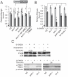
Figure 1. LRK-1 functions upstream of p38 to protect against 6-OHDA-induced DAergic neuron degeneration in C. elegans.
(A) Human LRRK2 can functionally substitute for LRK-1 to protect against 6-OHDA-induced DAergic neuron degeneration. Day 4 nematodes of the indicated genotypes (N2, three different lrk-1 loss-of-function mutants (km17, km41 and tm1898), and km17 mutants expressing human WT or K1906M (kinase inactive) LRRK2 in their DAergic neurons) were treated with vehicle (grey bars) or 2 mM 6-OHDA (black bars) for 1 h at L3. DAergic neurons were counted and normalized as described in Experimental Procedures. Data represent the mean ± SEM of three independent experiments. Each experiment employed 20–30 nematodes. #, p>0.05, *, p<0.05, **, p<0.01, and ***, p<0.005 by t-test. (B) lrk-1, pmk-1 and sek-1 function in the same genetic pathway to protect against 6-OHDA-induced DAergic neurodegeneration. Day 4 nematodes of the indicated genotypes (N2, km17 mutant (lrk-1), pmk-1/p38 null (pmk-1/p38), lrk-1pmk-1 double null (lrk-1/pmk-1) and sek-1/MKK6 null (sek-1)) were treated with vehicle (light grey bars) or 2 mM 6-OHDA (dark grey bars) for 1 h at L3. DAergic neurons were counted and normalized as described in Experimental Procedures. Data represent the mean ± SEM of four independent experiments. Each experiment employed 20–30 nematodes. *, p<0.05 and **, p<0.01 by one-way ANOVA with Dunnett's test. (C) 6-OHDA treatment induces p38 phosphorylation in nematodes (upper panel). Nematodes were treated with 2 mM 6-OHDA and 1 mM imipramine for 1 h at L3, and harvested after 15 mins or 24 hrs as indicated above each lane. Western blots were probed with antibodies specific to phosphorylated p38 (P-p38) or total p38. 6-OHDA-mediated induction of p38 phosphorylation in nematodes requires LRK-1 activity (lower panel). N2, pmk-1 mutant or lrk-1 mutant (km17) nematodes were exposed to 2 mM 6-OHDA for 1 hour at L3 and harvested after 4 hrs or 24 hrs. No p38 band was detected by Western blot in lysates from pmk-1/p38 null mutant animals.
Figure 2
Figure 2. LRRK2 signaling potentiates cell survival after 6-OHDA exposure.
(A) 6-OHDA induces two peaks of p38 phosphorylation and the later peak is 6-OHDA dose-dependent. Western blotting with antibodies against phosphorylated p38 (P-p38) and total p38. Lysates were prepared from SH-SY5Y cells treated with 100 µM 6-OHDA for the indicated times (upper panel) or with the indicated concentrations of 6-OHDA for 4 hours (lower panel). (B) 6-OHDA-induced p38 activation is dependent on LRRK2 function. Upper left panel: Western blot analysis of LRRK2 and β-tubulin (loading control) levels in parental SH-SY5Y cells (No KD), SH-SY5Y cells transfected with a control vector (vector), and SH-SY5Y cells from a MIX LRRK2 KD line (LRRK2 KD). Upper right panel: Western blot analysis of P-p38 and total p38 levels in SH-SY5Y cells transfected with a control vector (vector) or SH-SY5Y cells from a MIX LRRK2 KD line (LRRK2 KD) with or without exposure to 100 µM 6-OHDA for 4 hours. Lower panel: Western blot analysis of P-p38 and total p38 levels in SH-SY5Y cells transfected with a scramble shRNA (Scramble) or MIX LRRK2 shRNA (LRRK2 KD) with or without exposure to 100 µM 6-OHDA for 4 hours. (C) Expression of WT, but not K1906M mutant, LRRK2 restores 6-OHDA-induced p38 activation in LRRK2 KD cells. Western blot analyses of LRRK2, P-p38 and total p38 levels. Lysates were prepared from parental SH-SY5Y cells reconstituted with control vector (No KD)or 3′-UTR LRRK2 KD SH-SY5Y cells reconstituted with control vector (Control), K1906M or WT LRRK2 with or without exposure to 100 µM 6-OHDA for 4 hours. (D) Cells from a MIX LRRK2 KD SH-SY5Y line (LRRK2 KD, diamonds) and SH-SY5Y cells transfected with a control vector (Control, circles) were treated with 100 µM 6-OHDA for the indicated times. Cell viability was determined using an XTT-based calorimetric assay. Data represent the mean ± SEM of 3 independent experiments. **, p<0.01 by two-way ANOVA. (E) Cells with reduced LRRK2 expression show heightened sensitivity to 6-OHDA. Cells from a MIX LRRK2 KD SH-SY5Y line (LRRK2 KD, diamonds) and SH-SY5Y cells transfected with a control vector (Control, circles) were treated with the indicated concentrations of 6-OHDA for 24 hours. Cell viability was determined using an XTT-based calorimetric assay. Data represent the mean ± SEM of 3 (LRRK2 KD) or 4 (control) independent experiments. **, p<0.01 by two-way ANOVA. (F) Inhibition of p38 potentiates 6-OHDA-induced cell death in cells expressing LRRK2. SH-SY5Y cells transfected with control vector (Control) and MIX LRRK2 KD SH-SY5Y cells (LRRK2 KD) were treated with 100 µM 6-OHDA and either control vehicle or PD169316 for 24 hours. Cell survival was measured with a XTT-based calorimetric assay. Data represent the mean ± SEM of 4 independent experiments. ***, p<0.005 by t-test. (G) Expression of WT, but not kinase-inactive mutant, LRRK2 counteracts 6-OHDA-induced cell death. 3′-UTR LRRK2 KD SH-SY5Y cells reconstituted with K1906M mutant LRRK2 (diamonds) or WT LRRK2 (circles) were exposed to the indicated concentrations of 6-OHDA for 24 hours and cell survival was assessed with a XTT-based calorimetric assay. Data represent the mean ± SEM of 4 independent experiments. **, p<0.01 by two-way ANOVA, and *, P<0.05; **, P<0.01 by t-test.
Figure 3
Figure 3. LRRK2 signaling protects against hαSyn-mediated DAergic soma degeneration in C. elegans.
Quantification of DAergic neurons in nematodes of different genotypes with or without expression of human α-synuclein (hαSyn) in DAergic neurons. The number of DAergic somas was normalized to the mean number of DAergic somas observed in wild type (N2) L4 nematodes throughout this study, as described in Experimental Procedures. (A) Human α-synuclein induced death of DAergic neurons in nematodes is enhanced by mutations in components of the LRRK2 and p38 signaling pathway. Left panel, quantification of DAergic neuron degeneration as a function of age in Bristol N2 (N2, circles), loss-of-function mutant lrk-1 (km17) (triangles), human α-synuclein (hαSyn)-expressing (in DAergic neurons) N2 (hαSyn; N2, squares), and hαSyn-expressing lrk-1 (km17) (hαSyn; lrk-1(km17), diamonds) nematodes. The data shown represent the mean ± SEM of four independent experiments. Each experiment employed 20–30 nematodes. **, p<0.01 by two-way ANOVA. Right panel, quantification of DAergic neurons in wild type (Wild type) and different alleles of loss-of-function mutant lrk-1 (km17, tm1898 and km41) day 8 nematodes without (control, grey) or with (hαSyn, black) DAergic-specific expression of hαSyn. Data represent the mean ± SEM of four independent experiments. Each experiment employed 20–30 nematodes. **, p<0.01 by one-way ANOVA with Dunnett's test. For control (Wild type) and lrk-1 (km17) nematodes with and without hαSyn expression, the data in the right panel corresponds to that shown in the left panel for Day 8 (the same animals produced both data sets). (B) lrk-1 functions together with pmk-1 and sek-1 to protect against hαSyn-induced DAergic neurodegeneration. Quantification of DAergic neurons in wild type and the indicated single and double mutant day 8 nematodes without (control, grey) or with (black) DAergic-specific expression of hαSyn. Data represent the mean ± SEM of four independent experiments. Each experiment employed 20–30 nematodes. *, p<0.05; **, p<0.01; one-way ANOVA with Dunnett's test. (C) Human LRRK2 functionally substitutes for LRK-1 to protect nematode DAergic neurons from hαSyn-induced degeneration and LRRK2 kinase activity contributes to this protection. Quantification of DAergic neurons in day 8 nematodes including wild type (Wild type), lrk-1 mutant (km17), and km17 with pan-neuronal expression of WT (lrk-1+WT LRRK2) or K1906M mutant LRRK2 (lrk-1+K1906M LRRK2). All nematode strains also expressed hαSyn in their DAergic neurons. Data represent the mean ± SEM of four independent experiments. Each experiment employed 20–30 nematodes. #, p>0.05; *, p<0.05; **, p<0.01 by one-way ANOVA with Dunnett's test.
Figure 4
Figure 4. LRRK2 signaling modulates GRP78 levels to potentiate cell survival after 6-OHDA exposure.
(A) LRRK2 is involved in 6-OHDA-mediated upregulation of GRP78 protein levels. Western blot analyses were performed on lysates from SH-SY5Y cells transfected with a control vector (Control, circles) and MIX LRRK2 KD SH-SY5Y cells (LRRK2 KD, diamonds) treated with 100 µM 6-OHDA for the indicated periods of time. Representative Western blots are shown, as well as quantization of results from 3 independent experiments. Data represent the mean ± SEM of 3 independent experiments. **, p<0.01 by two-way ANOVA. (B) Induction of GRP78 via 6-OHDA/LRRK2 occurs at the mRNA level. qRT-PCR quantification of GRP78 and β-actin (housekeeping control for normalization) mRNA levels was performed on SH-SY5Y cells transfected with a control vector (circles) and MIX LRRK2 KD SH-SY5Y cells (squares) treated with 100 µM 6-OHDA for the indicated periods of time. Data represent the mean ± SEM of 3 independent experiments. **: p<0.01 by two-way ANOVA. (C) Inhibition of p38 with PD169316 compromises 6-OHDA-induced upregulation of GRP78. Western blotting was performed on SH-SY5Y cells treated with 100 µM 6-OHDA together with 20 µM PD169316 or vehicle for 8 hours. (D) LRRK2 kinase activity contributes to induction of GRP78 by 6-OHDA. Western blotting was performed on 3′-UTR LRRK2 KD SH-SY5Y cells transfected with WT (WT) or K1906M mutant LRRK2 (K1906M) treated with 100 µM 6-OHDA for 8 hours. Left panel, sample blots. Middle panel, quantification of Western blot results. Right panel, under similar experimental conditions, MIX LRRK2 KD SH-SY5Y cells (LRRK2 KD) displayed compromised 6-OHDA-mediated induction of GRP78 as compared to SH-SY5Y cells transfected with control vector (vector). Data in the middle and right panels represent the mean ± SEM of 3 independent experiments. ***, p<0.005 by t-test. (E) shRNA-mediated double KD of GRP78 and LRRK2 displayed a similar 6-OHDA-indcuced cell death rate to that observed in single KD of either GRP78 or LRRK2. Cell survival was assessed using an XTT-based calorimetric assay in Vector control SH-SY5Y cells transfected with a scramble shRNA (Control+Scramble), or GRP78 shRNA (Control+GRP78 KD) and in MIX LRRK2 KD SH-SY5Y cells transfected with scramble shRNA (LRRK2 KD+Scramble), or GRP78 shRNA (LRRK2 KD+GRP78 KD). Cells were exposed to 100 µM 6-OHDA for 12 hours prior to assessment of cell survival. Data are shown normalized to the level of survival in cell cultures not treated with 6-OHDA and represent the mean ± SEM of 3 independent experiments. ***, p<0.005; one-way ANOVA.
Figure 5
Figure 5. LRRK2 supports GRP78-mediated cell survival.
(A) Quantification of the populations of cells with sub-G0 DNA content in cultures of SH-SY5Y cells transfected with a control vector (Control) and MIX LRRK2 KD SH-SY5Y cells (LRRK2 KD) treated with 0, 3 or 6 µM tunicamycin for 16 hours. Data represent the mean ± SEM of 3 independent experiments. ***, p<0.005 by t-test. (B) Tunicamycin-induced upregulation of GRP78 is compromised in LRRK2 KD cells. SH-SY5Y cells transfected with a control vector (Control) and MIX LRRK2 KD SH-SY5Y cells (LRRK2 KD) were treated with 3 µM tunicamycin for the indicated periods of time. Cell lysates were prepared and analyzed by Western blotting with antibodies against GRP78 and β-tubulin (loading control). (C) 6-OHDA induced HSP-4::GFP upregulation in N2, but not in the lrk-1 mutant. L3 animals were treated with 2 mM 6-OHDA or vehicle alone (Control) for 1 h. Twenty-four hrs later, these animals were immobilized on 2% agarose pads with 3 mM sodium azide and then examined under a Leica DMI3000 microscope. HSP-4::GFP fluorescence of each animal was quantified as described in Experimental Procedures. Data represent the mean ± SEM. Each experiment employed 20 nematodes. *, p<0.05 by t-test. (D) Day 6 aniamls of the indicated genotypes were treated with vehicle alone (Control) or 2 mM 6-OHDA (6-OHDA) for 1 h at L3. DAergic neurons were counted and normalized as described in Experimental Procedures. Data represent the mean ± SEM of three independent experiments. Each experiment employed 20–30 nematodes. #, p>0.05 by one-way ANOVA with Bonferroni correction, and *, p<0.05; **, p<0.01; ***, P<0.005 by t-test. (E) HSP-4 protects hαSyn-induced DAergic neuron degeneration in C. elegans. Quantification of hαSyn-induced DAergic neuron degeneration as a function of age in N2 (squares) and loss-of-function hsp-4 mutant nematodes (circles). An hsp-4 nematode line expressing only DsRed served as a control (diamonds). DAergic neuron somas of hermaphrodites were counted on the indicated days and normalized to the mean number of DAergic somas observed in respective L4 nematode larvae as described in Experimental Procedures. Data represent the mean ± SEM of three independent experiments. Each experiment employed 40–60 nematodes. **, p<0.01 by two-way ANOVA, testing samples between hsp-4 and N2 nematodes expressing hαSyn.
Figure 6
Figure 6. Expression of human pathogenic G2019S mutant LRRK2 in nematodes leads to ageing-associated DAergic-specific neurodegeneration and chronic p38 activation.
(A) Quantification of DAergic neurons as a function of nematode age demonstrates that G2019S expression in nematodes elicited LRRK2 kinase-dependent DAergic neuron degeneration. Nematode variants were N2 nematodes (diamonds), N2 nematodes expressing K1906M LRRK2 (triangles), WT LRRK2 (squares), or G2019S LRRK2 (crosses) and lrk-1 mutant nematodes (km17) expressing G2019S LRRK2 (circles). The number of DAergic somas was normalized to the mean number of DAergic somas observed in N2 L4 nematodes throughout the study as described in Experimental Procedures. Data represent the mean ± SEM of four independent experiments. Each experiment employed 20–30 nematodes. ***, p<0.005; two-way ANOVA compared N2 nematodes expressing G2019S mutant LRRK2 with N2 nematodes expressing WT LRRK2. (B) G2019S-mediated neurodegeneration in nematodes is DAergic-specific. DAergic neuron and command interneuron somas were quantified in day 8 N2 nematodes with (black) or without (grey) pan-neuronal expression of G2019S mutant LRRK2. Somas of DAergic neurons and command interneurons were visualized with Pdat-1 driven DsRed or Pnmr-1 driven YFP, respectively. Command interneurons were not DAergic. Only AVA and PVC command interneurons were used for this quantification because their fluorescent somas can be unambiguously distinguished from other command interneurons in nematodes due to their position and stronger YFP expression. The numbers of DAergic or command interneuron somas were normalized to the mean number of DAergic or command interneuron somas observed in N2 L4 nematodes, respectively (see Experimental Procedures). Data represent the mean ± SEM of four (DAergic neuron) or three (command interneuron) independent experiments. 20–30 nematodes were used for each experiment. ***, p<0.005 by t-test. (C) LRRK2 KD cells reconstituted with G2019S LRRK2 have a high basal level of activated p38, but are comparable to cells reconstituted with WT LRRK2 in their response to 6-OHDA. Western blotting was performed using lysates from 3′-UTR LRRK2 KD SH-SY5Y cells expressing G2019S mutant LRRK2 (G2019S) or WT LRRK2 (WT). Cells were left untreated or treated with 100 µM 6-OHDA for 4 hours. (D) p38 prevents G2019S-mediated adult onset DAergic neurodegeneration in nematodes. DAergic neuron somas were counted in L4 (day 0, grey) or day 8 (black) N2 or pmk-1 mutant nematodes with pan-neuronal G2019S expression. Data was normalized to respective day 0 animals and represent the mean ± SEM of 3 independent experiments (20–30 nematodes each). ***, p<0.005 by t-test.
Figure 7
Figure 7. Activation of p38 contributes to cytotoxicity induced by G2019S mutant LRRK2 expression in murine neurons.
Murine primary cortical neurons transfected with empty vector (control), K1906M or G2919S LRRK2 along with pEGFP vector were left untreated (vehicle) or incubated with 20 µM PD169316 for two days followed by TUNEL assay (a–b) or viability assay (C–D). (A, C) Representative fluorescent microscopic images of TUNEL assay (A) or viability assay (C). BF, bright field images; merged, merged images of BF, GFP and TUNEL. (B, D) Quantification of neuron apoptosis by TUNEL assay (B) or of neuron viability (D) demonstrates that p38 inhibition decreases the cytotoxicity of G2019S LRRK2 expressed in murine primary cortical neurons. For quantification of apoptotic cells, GFP- and TUNEL-positive neurons were counted as a percentage of total GFP-positive neurons in 40× microscopic fields. Percentages of TUNEL- positive control vector transfected neurons with or without PD169316 treatment were subtracted from the respective percentages of neurons transfected with other constructs. In viability assays, GFP-positive neurons with neurites (See Experimental Procedures) were counted as a percentage of total GFP-positive neurons. Data were normalized to neurons transfected with control vector. Data represent the mean ± SEM of three independent experiments. * p<0.05 by t-test.

Similar articles

Cited by

References

    1. Zimprich A, Biskup S, Leitner P, Lichtner P, Farrer M, et al. Mutations in LRRK2 cause autosomal-dominant parkinsonism with pleomorphic pathology. Neuron. 2004;44:601–607. - PubMed
    1. Paisan-Ruiz C, Jain S, Evans EW, Gilks WP, Simon J, et al. Cloning of the gene containing mutations that cause PARK8-linked Parkinson's disease. Neuron. 2004;44:595–600. - PubMed
    1. Paisan-Ruiz C. LRRK2 gene variation and its contribution to Parkinson disease. Hum Mutat. 2009;30:1153–1160. - PubMed
    1. West AB, Moore DJ, Biskup S, Bugayenko A, Smith WW, et al. Parkinson's disease-associated mutations in leucine-rich repeat kinase 2 augment kinase activity. Proc Natl Acad Sci U S A. 2005;102:16842–16847. - PMC - PubMed
    1. Lee BD, Shin JH, Vankampen J, Petrucelli L, West AB, et al. Inhibitors of leucine-rich repeat kinase-2 protect against models of Parkinson's disease. Nat Med 2010 - PMC - PubMed

Publication types

MeSH terms

Substances