. 2011;6(8):e23094.
doi: 10.1371/journal.pone.0023094. Epub 2011 Aug 8.

Increased muscle stress-sensitivity induced by selenoprotein N inactivation in mouse: a mammalian model for SEPN1-related myopathy

Affiliations

Increased muscle stress-sensitivity induced by selenoprotein N inactivation in mouse: a mammalian model for SEPN1-related myopathy

Mathieu Rederstorff et al. PLoS One. 2011.

Abstract

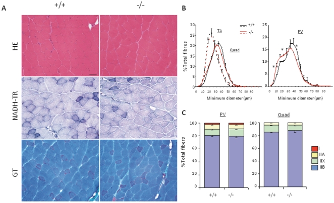
Selenium is an essential trace element and selenoprotein N (SelN) was the first selenium-containing protein shown to be directly involved in human inherited diseases. Mutations in the SEPN1 gene, encoding SelN, cause a group of muscular disorders characterized by predominant affection of axial muscles. SelN has been shown to participate in calcium and redox homeostasis, but its pathophysiological role in skeletal muscle remains largely unknown. To address SelN function in vivo, we generated a Sepn1-null mouse model by gene targeting. The Sepn1(-/-) mice had normal growth and lifespan, and were macroscopically indistinguishable from wild-type littermates. Only minor defects were observed in muscle morphology and contractile properties in SelN-deficient mice in basal conditions. However, when subjected to challenging physical exercise and stress conditions (forced swimming test), Sepn1(-/-) mice developed an obvious phenotype, characterized by limited motility and body rigidity during the swimming session, as well as a progressive curvature of the spine and predominant alteration of paravertebral muscles. This induced phenotype recapitulates the distribution of muscle involvement in patients with SEPN1-Related Myopathy, hence positioning this new animal model as a valuable tool to dissect the role of SelN in muscle function and to characterize the pathophysiological process.


Conflict of interest statement

Competing Interests: The authors have declared that no competing interests exist.

Figures

Figure 1
Figure 1. Generation of the Sepn1 knock-out mouse model.
A) Strategy for the selective excision of Sepn1 exon 3. In the targeting fragment, LoxP sites were incorporated on each side of exon 3 together with a floxed neomycin (neo) cassette (L3). This fragment was electroporated into ES cells and neo-resistant clones that featured homologous recombination were selected. In vivo or ex vivo Cre-induced excision of floxed regions leading to neo excision (L2) or complete excision (L-) are represented. B) Mice genotyping. PCR were performed on genomic DNA extracted from Sepn1+/+, Sepn1+/− and Sepn1−/− mice. Exon 3 excision leads to a 500 bp loss in the PCR product size. The bands at 1000 and 490 bp represent the wild-type and the exon 3-deleted alleles, respectively. C) Expression of Sepn1 transcript in Sepn1+/+, Sepn1+/− and Sepn1−/− tissues. Brain, quadriceps (quad) and diaphragm (diaph) were tested. Quantification was performed by qRT-PCR and normalized to the 18S gene expression. n = 3, *, p<0.05; **, p<0.01. D) Selenoprotein N (SelN) expression. Western blot analysis was performed using total protein extracts from several tissues (Sk M, skeletal muscle) of Sepn1−/− and Sepn1+/+ mice. GAPDH was used as a loading control. E) Body weight analysis. Body weight evolution of wild-type or mutant, male or female mice, from 5 to 40 weeks of age was monitored. Similar growth curves were observed for mutant and control mice of both genders.
Figure 2
Figure 2. Histomorphology of Sepn1−/− adult muscles.
A) Representative image of quadriceps cryosections, stained with HE, NADH-TR and GT, from 10 month-old mutant and control mice. Mutant muscle displayed no obvious histopathological abnormalities. Scale bar, 50 µm. B) Fiber size distribution (% of total number of fibers) according to their minimum diameter (µm) for TA and quadriceps muscles (left panel) and paravertebral (PV) muscles (right panel) from 10 month-old mutant and wild-type mice. A switch toward smaller fibers was observed in Sepn1−/− PV muscles compared to wild-types. n = 3, * p<0.05; ** p<0.001. C) Fiber type distribution (% of total number of fibers) based on type I and II MHC immunostaining of paravertebral and quadriceps muscles, from 10 month-old mice. No significant difference was found between mutant and control mice. n = 3. Data are means±SEM.
Figure 3
Figure 3. Ultrastructural organization and triadic junctions in Sepn1−/− adult muscles.
A) Electron microscopy image of gastrocnemius muscles from 6 month-old wild-type and mutant mice. No defect in sarcomere organization, mitochondria morphology (arrows) and triads structure (brackets) were observed in mutant (right panel), compared to wild-types (left panel). Scale bars, 1 µm (top) and 100 nm (bottom). B) Immunostaining for DHPR (green) and RyR1 or RyR3 (red) on longitudinal sections of quadriceps from 2 month-old mice, revealing normal localization of both receptors in mutant mice compared to controls. Nuclei are revealed by DAPI staining. Boxed regions in the merged images are magnified as inserts (right) showing intracellular co-localization (yellow). Scale bar is 10 µm. C) Skeletal muscle microsomes extracts from Sepn1 +/+ or Sepn1 −/− littermates were immunoblotted for Ryanodine Receptor type 1 (RyR1), Ryanodine Receptor type 3 (RyR3), Dihydropyridine Receptor (DHPR), Selenoprotein N (SelN) and triadin isoform Trisk 95 (Trisk 95).
Figure 4
Figure 4. Global phenotype of Sepn1−/− mice submitted to repeated forced swimming tests (FST).
A) Global appearance of 8 and 14 month-old wild-type and mutant mice, after 3 months of FST. A severe kyphosis was observed in both young and older mutant mice, while no sign of kyphosis was present in wild-type animals. B) Sagittal tomography of wild-type and mutant mice placed in ventral position, indicating a marked axial distortion of the spine in Sepn1 −/− animals (arrows). C) Gain of body mass for wild-type and mutant mice, after 1.5 and 3 months of FST. The gain was significantly lower in mutant mice, compared to wild-types, at both stages. n = 20 Sepn1 +/+ vs. 22 Sepn1 −/− for 1.5 months, n = 14 Sepn1 +/+ vs. 22 Sepn1 −/− for 3 months. * p<0.01, ** p<0.0005. D) Muscle masses normalized to body weight (%) in wild-type and mutant mice after 3 months of FST. Atrophy was observed for TA, quadriceps (Quad), gastrocnemius/plantaris/soleus muscles group (GPS) and extensor digitorum longus (EDL) muscles from mutant mice, compared to wild-types. Diaphragm (Diaph) and heart masses were not affected. n = 11 Sepn1 +/+ vs. 16 Sepn1 −/− (except for diaphragm: n = 8 Sepn1 +/+ vs. 10 Sepn1 −/−), * p<0.05, ** p<0.001, *** p<0.0005, **** p<0.0001. Data are means±SEM.
Figure 5
Figure 5. Histomorphology of Sepn1−/− muscles after 3 months of FST.
A) Representative images of quadriceps sections, stained with HE, NADH-TR and GT, from 8 and 14 month-old mice submitted to 3 months of FST, as indicated. Cytoplasmic inclusions, stained in red by GT (arrows), were detected in several muscle fibers from young mice, and were much more abundant in old mutant mice. Aggregates were only occasionally observed in Sepn1 +/+ mice. Scale bar, 50 µm. B) Representative images of tubular aggregates observed in mutant quadriceps (left panel) and gastrocnemius (right panel) from old mutant mice, after FST, by electron microscopy. Accumulation of sarcoplasmic reticulum membranes was observed in the majority of mutant fibers, leading to large aggregates inclusions in the cytoplasm. Scale bar, 1 µm. C) Fiber size distribution, according to their minimum diameter (µm), for TA and quadriceps muscles (right panel) and paravertebral muscles (left panel) from 8 month-old mice, at the end of the test. Selective specific switch towards smaller fibers was observed in mutant paravertebral muscles, while no major alteration was detected for other muscles. n = 4 Sepn1 +/+ vs. 6 Sepn1 −/−, * p<0.05. D) Fiber type repartition (% of total number of fibers) based on type I and II MHC immunostaining for paravertebral and quadriceps muscles, from 8 month-old mice, after FST. Significant switch from type IIB to types IIX and IIA was only observed for paravertebral muscles in mutant mice, compared to wild-types. n = 4 Sepn1 +/+ vs. 6 Sepn1 −/−, * p<0.005, ** p<0.001. Data are means±SEM.

Similar articles

Cited by

References

    1. Rayman MP. Selenoproteins and human health: insights from epidemiological data. Biochim Biophys Acta. 2009;1790:1533–1540. - PubMed
    1. Hatfield DL, Yoo MH, Carlson BA, Gladyshev VN. Selenoproteins that function in cancer prevention and promotion. Biochim Biophys Acta. 2009;1790:1541–1545. - PMC - PubMed
    1. Allmang C, Wurth L, Krol A. The selenium to selenoprotein pathway in eukaryotes: more molecular partners than anticipated. Biochim Biophys Acta. 2009;1790:1415–1423. - PubMed
    1. Arner ES. Selenoproteins-What unique properties can arise with selenocysteine in place of cysteine? Exp Cell Res. 2010;316:1296–1303. - PubMed
    1. Forman HJ, Maiorino M, Ursini F. Signaling functions of reactive oxygen species. Biochemistry. 2010;49:835–842. - PMC - PubMed

Publication types

MeSH terms