. 2011;6(8):e23139.
doi: 10.1371/journal.pone.0023139. Epub 2011 Aug 9.

Contrasting expression of canonical Wnt signaling reporters TOPGAL, BATGAL and Axin2(LacZ) during murine lung development and repair

Affiliations

Contrasting expression of canonical Wnt signaling reporters TOPGAL, BATGAL and Axin2(LacZ) during murine lung development and repair

Denise Al Alam et al. PLoS One. 2011.

Abstract

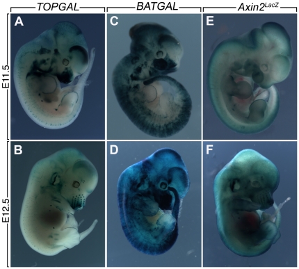
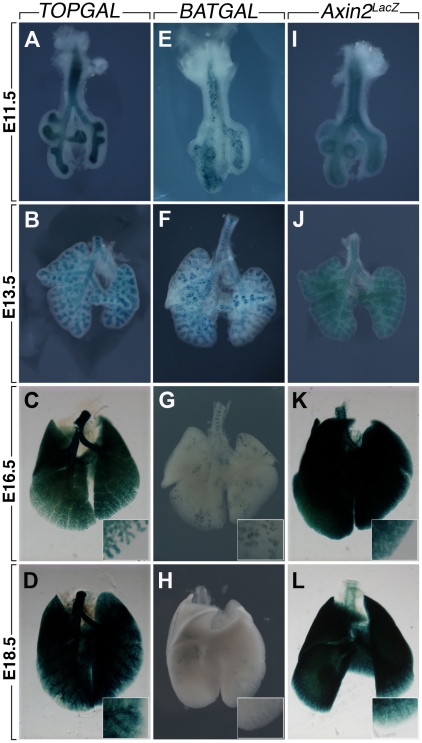
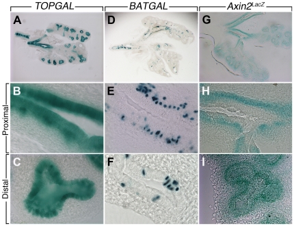
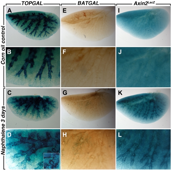
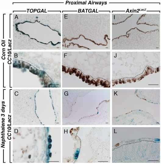
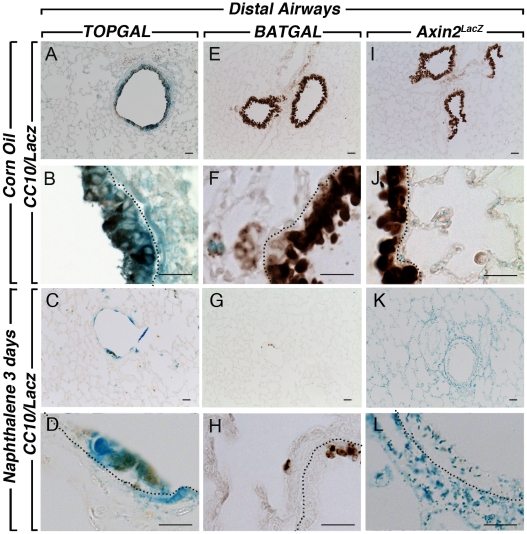
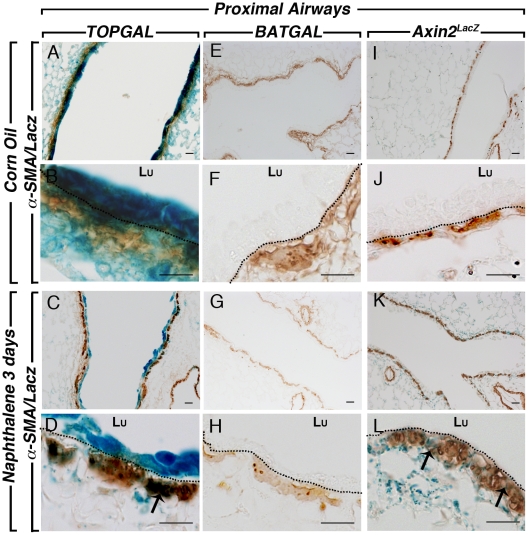
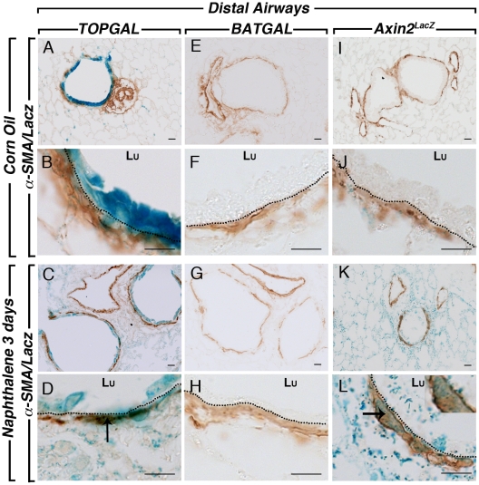
Canonical WNT signaling plays multiple roles in lung organogenesis and repair by regulating early progenitor cell fates: investigation has been enhanced by canonical Wnt reporter mice, TOPGAL, BATGAL and Axin2(LacZ). Although widely used, it remains unclear whether these reporters convey the same information about canonical Wnt signaling. We therefore compared beta-galactosidase expression patterns in canonical Wnt signaling of these reporter mice in whole embryo versus isolated prenatal lungs. To determine if expression varied further during repair, we analyzed comparative pulmonary expression of beta-galactosidase after naphthalene injury. Our data show important differences between reporter mice. While TOPGAL and BATGAL lines demonstrate Wnt signaling well in early lung epithelium, BATGAL expression is markedly reduced in late embryonic and adult lungs. By contrast, Axin2(LacZ) expression is sustained in embryonic lung mesenchyme as well as epithelium. Three days into repair after naphthalene, BATGAL expression is induced in bronchial epithelium as well as TOPGAL expression (already strongly expressed without injury). Axin2(LacZ) expression is increased in bronchial epithelium of injured lungs. Interestingly, both TOPGAL and Axin2(LacZ) are up regulated in parabronchial smooth muscle cells during repair. Therefore the optimal choice of Wnt reporter line depends on whether up- or down-regulation of canonical Wnt signal reporting in either lung epithelium or mesenchyme is being compared.


Conflict of interest statement

Competing Interests: The authors have declared that no competing interests exist.

Figures

Figure 1
Figure 1. LacZ expression in whole embryos of TOPGAL, BATGAL and Axin2LacZ mice.
(A) E11.5 TOPGAL embryo shows staining in the forebrain, the nasal process, the inner ear, the apical ectodermal ridge (AER) of the limb, the epithelium of the mammary placode, the somites and the tip of the tail. (B) E12.5 TOPGAL embryo shows LacZ expression in ectodermal appendages, the whisker placodes in the nasal region as well as the AER, the mammary buds in between the limbs and discrete mesenchymal condensation within the limbs. (C) E11.5 BATGAL embryo shows expression throughout the embryo with a “salt and pepper” pattern with higher signals in ectodermal domains such as the nasal process, the forebrain, the AER and the tip of the tail. (D) At E12.5, the BATGAL embryo still shows “salt and pepper” expression throughout the embryo. LacZ expression is found in the mammary buds but significant staining was also found in the surrounding tissue. (E) E11.5 Axin2LacZ embryo shows homogenous LacZ expression throughout the embryo with higher expression in the AER and the developing mammary placode. (F) At E12.5, individualized whisker placodes as well as mammary buds and the AER are clearly positive for Axin2LacZ. In addition, strong LacZ expression is found in the developing ear and in the somites.
Figure 2
Figure 2. LacZ expression in TOPGAL, BATGAL and Axin2LacZ whole lungs during prenatal development.
TOPGAL is expressed in the epithelium of the trachea and the lung at E11.5 (A), E13.5 (B) and E16.5 (C); whereas at E18.5 (D) TOPGAL expression is also detected in the terminal bronchioles and the surrounding parenchyma. BATGAL expression is detected in a “salt and pepper” manner in the lung epithelium at E11.5 (E) and E13.5 (F). This expression is drastically reduced at E16.5 (G) and E18.5 (H). Axin2LacZ expression is found in both the epithelium and mesenchyme at E11.5 (I), E13.5 (J), E16.5 (K) and e18.5 (L).
Figure 3
Figure 3. Vibratome sections of E13.5 TOPGAL, BATGAL and Axin2LacZ lungs.
TOPGAL is expressed in both epithelium and mesenchyme at the level of the bronchi (A, B) and restricted to the epithelium in the distal lung (C). BATGAL “salt and pepper” expression is found in a heterogeneous fashion in the lung (D). The expression is restricted to the mesenchyme adjacent to the bronchial epithelium (E). In the distal lung, BATGAL is sporadically expressed in the epithelium (F). Axin2LacZ is found exclusively in the mesenchyme adjacent to the epithelium at the bronchial level (G, H). While in the distal lung, LacZ expression is found mainly in the epithelium and at lower level in the mesenchyme (I).
Figure 4
Figure 4. Corn oil and Naphthalene-treated whole mount left lungs cleared with Benzyl benzoate.
In the adult lung, TOPGAL is strongly expressed in the epithelium (A, B) and decreases slightly after naphthalene injury (C, D). BATGAL expression is totally absent in the adult lung (E, F) and a discrete expression appears after injury (G, H). While a homogenous Axin2LacZ expression is present throughout the adult lung (I, J), a marked increase surrounding the bronchial epithelium is observed after injury (K, L).
Figure 5
Figure 5. CC10 and LacZ staining in the proximal lungs after naphthalene injury.
CC10 and LacZ co-staining in TOPGAL control lungs at low (A) and high (B) magnification show strong expression of TOPGAL in the CC10-positive cells. A decrease in the Clara cells after naphthalene injury is shown in the proximal airways (C, D). BATGAL expression is not detected in the airways (E, F) but at discrete spots in the bronchial epithelium after injury (G, H). Axin2LacZ sections showed very low level staining throughout the lung in the conducting airways of the control adult lungs (I, J), and an increased expression after injury (K, L). Scale bars are 100 µm.
Figure 6
Figure 6. CC10 and LacZ staining in the distal lung compartment after naphthalene injury.
CC10 and LacZ co-staining in TOPGAL control lungs at low (A) and high (B) magnification show strong expression of TOPGAL in the CC10-positive cells. A decrease in the Clara cells after naphthalene injury is shown in the distal airways (C, D). BATGAL expression is not detected in the distal airways (E, F) of adult control lungs, and still absent from the epithelium (G, H) in the distal compartment of the naphthalene-treated lungs. Axin2LacZ sections showed very low level staining in the distal airways of the control adult lungs (I, J), and an increased expression after injury (K, L). Scale bars are 100 µm.
Figure 7
Figure 7. SMA and LacZ staining in the proximal lung compartment after naphthalene injury.
Co-staining of SMA and LacZ at low (A) and high (B) magnifications showed co-localization in the PBSMCs of control and naphthalene-treated lungs (C, D). BATGAL expression is not detected in the proximal PBSMCs in the control lung (E, F) and naphthalene-treated lungs (G, H). Axin2LacZ sections showed low level staining in the PBMSCs of the control adult lungs (I, J) whereas LacZ expression is drastically upregulated after injury as pointed out with the arrows (K, L). Dotted lines show the basal membrane separating the epithelium from the PSMC layer. Scale bars are 100 µm.
Figure 8
Figure 8. SMA and LacZ staining in the distal lung compartment after naphthalene injury.
SMA and LacZ co-staining in TOPGAL control lungs at low (A) and high (B) magnification showed co-localization in the PBSMCs of control (A, B) and naphthalene-treated lungs (C, D). BATGAL expression is not detected in the PBSMCs of adult control lungs (E, F) and naphthalene-treated lungs (G, H). Axin2LacZ sections showed low level staining in the PBSMCs surrounding the distal bronchioles in control adult lungs (I, J) and an increase after injury (K, L). The arrows show co-localisation of SMA and LacZ. Dotted lines show the basal membrane separating the epithelium from the PSMC layer. Scale bars are 100 µm.

Similar articles

Cited by

References

    1. De Langhe SP, Reynolds SD. Wnt signaling in lung organogenesis. Organogenesis. 2008;4:100–108. - PMC - PubMed
    1. Warburton D, El-Hashash A, Carraro G, Tiozzo C, Sala F, et al. Lung organogenesis. Curr Top Dev Biol. 90:73–158. - PMC - PubMed
    1. Nusse R. WNT targets. Repression and activation. Trends Genet. 1999;15:1–3. - PubMed
    1. Brembeck FH, Schwarz-Romond T, Bakkers J, Wilhelm S, Hammerschmidt M, et al. Essential role of BCL9-2 in the switch between beta-catenin's adhesive and transcriptional functions. Genes Dev. 2004;18:2225–2230. - PMC - PubMed
    1. Price MA. CKI, there's more than one: casein kinase I family members in Wnt and Hedgehog signaling. Genes Dev. 2006;20:399–410. - PubMed

Publication types

MeSH terms

LinkOut - more resources