. 2011;6(8):e23246.
doi: 10.1371/journal.pone.0023246. Epub 2011 Aug 17.

The kinase inhibitor SFV785 dislocates dengue virus envelope protein from the replication complex and blocks virus assembly

Affiliations

The kinase inhibitor SFV785 dislocates dengue virus envelope protein from the replication complex and blocks virus assembly

Azlinda Anwar et al. PLoS One. 2011.

Abstract

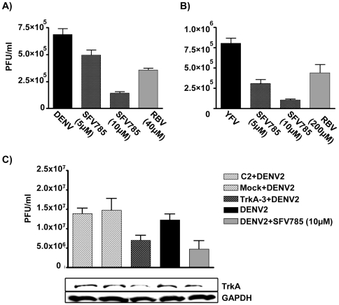
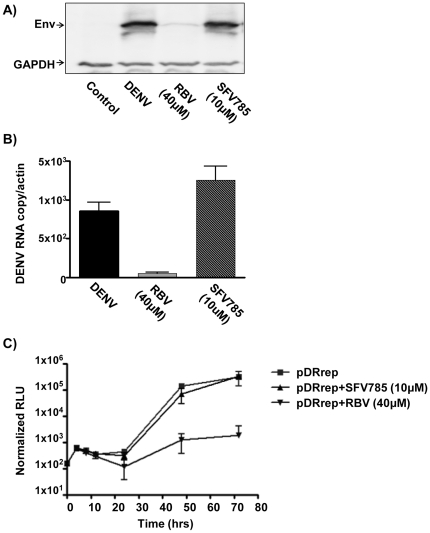
Dengue virus (DENV) is the etiologic agent for dengue fever, for which there is no approved vaccine or specific anti-viral drug. As a remedy for this, we explored the use of compounds that interfere with the action of required host factors and describe here the characterization of a kinase inhibitor (SFV785), which has selective effects on NTRK1 and MAPKAPK5 kinase activity, and anti-viral activity on Hepatitis C, DENV and yellow fever viruses. SFV785 inhibited DENV propagation without inhibiting DENV RNA synthesis or translation. The compound did not cause any changes in the cellular distribution of non-structural 3, a protein critical for DENV RNA synthesis, but altered the distribution of the structural envelope protein from a reticulate network to enlarged discrete vesicles, which altered the co-localization with the DENV replication complex. Ultrastructural electron microscopy analyses of DENV-infected SFV785-treated cells showed the presence of viral particles that were distinctly different from viable enveloped virions within enlarged ER cisternae. These viral particles were devoid of the dense nucleocapsid. The secretion of the viral particles was not inhibited by SFV785, however a reduction in the amount of secreted infectious virions, DENV RNA and capsid were observed. Collectively, these observations suggest that SFV785 inhibited the recruitment and assembly of the nucleocapsid in specific ER compartments during the DENV assembly process and hence the production of infectious DENV. SFV785 and derivative compounds could be useful biochemical probes to explore the DENV lifecycle and could also represent a new class of anti-virals.


Conflict of interest statement

Competing Interests: Some of the authors have an ownership stake in the company KinoPharma. This does not alter the authors' adherence to all the PLoS ONE policies on sharing data and materials.

Figures

Figure 1
Figure 1. Structure-activity relationship of SRPIN340/nicotinoyl derivatives to the replication of the HCV subreplicon.
The comparison of the relative luciferase activity (luc activity) and viability (viability) with the solvent (0.04% DMSO) in the presence of 10 or 20 µM of the compounds are shown. L/V is the ratio of relative luciferase activity to viability. L/V values less than 50 (20 µM) or 60 (10 µM) are highlighted. The data are represented as the mean of triplicate experiments.
Figure 2
Figure 2. SFV785 inhibits HCV replication.
(A) Detection of HCV JFH1-infected cells. HuH 7.5-1 cells were infected with HCV JFH1 and treated with the compounds SFV785 or SRPIN614 for 48 hours. The cells were fixed and immunostained with an anti-HCV NS3 antibody. (B) Quantitative analysis of HCV JFH1-infected cells. HuH-7.5-1 cells were seeded in 96 well plates, infected with HCV JFH1 and treated with the compounds SFV785 or SRPIN614 as described above. The fixed cells were immunostained with an anti-HCV NS3 mouse monoclonal antibody, and the percentage of NS3-positive cells measured using the ArrayScan VTi. The bars indicate the mean value ± S.D. from triplicate experiments.
Figure 3
Figure 3. SFV785 inhibits DENV-2 and YFV propagation.
(A) DENV anti-viral activity of SFV785. HuH-7 cells were infected with DENV and treated with SFV785 (5 or 10 µM) or with ribavirin (40 µM). The culture media were collected 36 hrs post-infection (p.i) and measured for viral titer by plaque assays. (B) HuH-7 cells were infected with YFV and treated with SFV785 (5 or 10 µM) or ribavirin (200 µM). The culture media were collected 12 hrs p.i and assessed by plaque assays for viral titer. The bars indicate the range of PFU/ml from three biological replicates. (C) Knockdown of NTRK1, one of the targets of SFV785, inhibited DENV propagation. The culture media and cell lysates from DENV infected HuH-7 cells treated with mock transfection, C2 or NTRK1 (TrkA-3) siRNAs, were collected 36 hrs p.i. and analysed for viral titer by plaque assays and Western blot analysis, respectively. Cells treated with SFV785 (10 µM) and infected with DENV were used as a control. The bars indicate the range of PFU/ml from three biological replicates, each in triplicates. Similar results were obtained when NTRK1 knockdown was performed using TrkA-12 siRNA.
Figure 4
Figure 4. SFV785 did not inhibit DENV viral RNA synthesis or translation.
HuH-7 cells were infected with DENV, and untreated or treated with SFV785 (10 µM) or with ribavirin (40 µM). The levels of (A) DENV protein in the cellular lysates by Western blot analysis, and (B) DENV RNA by real-time PCR, are shown. The bars indicate the range of DENV copy number, normalized to cellular actin, from three biological replicates, each in triplicates. (C) Effect on SFV785 on the replication of the DENV replicon. HuH-7 cells electroporated with the DENV replicons were pooled and aliquoted in media untreated or treated with SFV785 (10 µM) or ribavirin (40 µM). The cells were harvested at the indicated time points and the lysates assessed for luciferase activity. The bars indicate the S.D. from 4 separate experiments.
Figure 5
Figure 5. The distribution profile of the dengue viral Env, but not the NS3, is altered in infected cells treated with SFV785, and does not co-localize with the replication complex.
HuH-7 cells were infected with DENV and were either untreated or treated with SFV785 (10 µM) for 36 hrs. The distribution profiles of the DENV (A) Env and (B) NS3 proteins were visualized by fluorescent microscopy. (C) DENV Env (green) and dsRNA (red) were visualized by fluorescent microscopy. Arrowheads indicate representative cells showing no co-localization of the vescicular-distributed Env with the dsRNA, a presumed marker for the DENV RNA replication. The nuclei in all experiments were stained with DAPI (blue).
Figure 6
Figure 6. Intracellular DENV protein is located within the endoplasmic reticulum in SFV785-treated cells.
The subcellular localization of DENV Env (green) proteins in DENV-infected HuH-7 cells, untreated or treated with SFV785 (10 µM), was visualized by fluorescent microscopy, with the nuclei stained with DAPI (blue). Antibodies used for the detection of cellular marker proteins are indicated in the respective panels (red). The cellular markers used were (A) calnexin (CALN) for the endoplasmic reticulum (ER), (B) ERGIC for the ER-Golgi intermediate compartments, (C) SEC31 for the COPII vesicles, and (D) GOLGB1 for the Golgi.
Figure 7
Figure 7. Ultrastructural analysis of DENV-infected or uninfected HuH-7 cells that were either untreated or treated with SFV785.
HuH-7 cells were mock-infected (A, B) or infected with DENV (C–F) at an MOI of 1, and either untreated (A, C, E) or treated with SFV785 (10 µM) (B, D, F). Cells were fixed at the late cycle of infection, incubated with 4% paraformaldehyde-PBS solution, and processed and analyzed by electron microscopy. CM, convoluted membranes; D, probable defective virus; ER, endoplasmic reticulum; M, mitochondria; N, nucleus; T, virus-induced tubules; Vd, probable drug-induced vacuoles; Ve, virus-induced vesicles; Vi, stacked DENV particles.
Figure 8
Figure 8. SFV785 induces the production of non-infectious virus lacking the nucleocapsid, without affecting viral secretion.
HuH-7 cells were infected with DENV and either untreated or treated with SFV785 (10 µM) or with ribavirin (40 µM). The culture media were collected 36 hrs p.i., and measured for (A) the titers of infectious DENV produced by plaque assay and (B) the supernatant viral RNA. The bars indicate the S.D. from at least 3 separate experiments. (C) Comparison of the concomitant levels of DENV protein from the supernatants of treated cells in (A). The amount of DENV proteins was quantified by antigen-capture ELISA and shown as a percentage of the DENV proteins in the control untreated (DENV) supernatant. The bars indicate the range of DENV proteins from triplicates of 2 independent experiments. P values indicate significant difference by 1-way ANOVA and Tukey's post-test. (D) Western blot analysis of the capsid (C) and pre-membrane (prM) structural proteins from ultracentifugation-concentrated supernatants of HuH-7 cells that were uninfected (control) or infected with DENV-2 and treated with or without SFV785.

Similar articles

Cited by

References

    1. Halstead SB. Dengue. Lancet. 2007;370:1644–1652. - PubMed
    1. Lindenbach BD, Thiel HJ, Rice CM. Flaviviridae: The Viruses and Their Replication. In: Knipe DM, Howley PM, editors. Fields Virology. 5th ed. Philadelphia: Lippincott-Raven Publishers; 2007. pp. 1101–1152.
    1. Webster DP, Farrar J, Rowland-Jones S. Progress towards a dengue vaccine. Lancet Infect Dis. 2009;9:678–687. - PubMed
    1. Pediatric Dengue Vaccine Initiative, Global burden of dengue, International Vaccine Institute http://www.pdvi.org/about_dengue/GBD.asp 30 Mar 2011.
    1. Welsch S, Miller S, Romero-Brey I, Merz A, Bleck CK, et al. Composition and three-dimensional architecture of the dengue virus replication and assembly sites. Cell Host Microbe. 2009;5:365–375. - PMC - PubMed

Publication types

MeSH terms

Substances