. 2011;6(8):e23268.
doi: 10.1371/journal.pone.0023268. Epub 2011 Aug 9.

Adverse effects of simulated hyper- and hypo-phosphatemia on endothelial cell function and viability

Affiliations

Adverse effects of simulated hyper- and hypo-phosphatemia on endothelial cell function and viability

Ai Peng et al. PLoS One. 2011.

Abstract

Background: Dysregulation of phosphate homeostasis as occurs in chronic kidney disease is associated with cardiovascular complications. It has been suggested that both hyperphosphatemia and hypophosphatemia can cause cardiovascular disease. The molecular mechanisms by which high or low serum phosphate levels adversely affect cardiovascular function are poorly understood. The purpose of this study was to explore the mechanisms of endothelial dysfunction in the presence of non-physiologic phosphate levels.

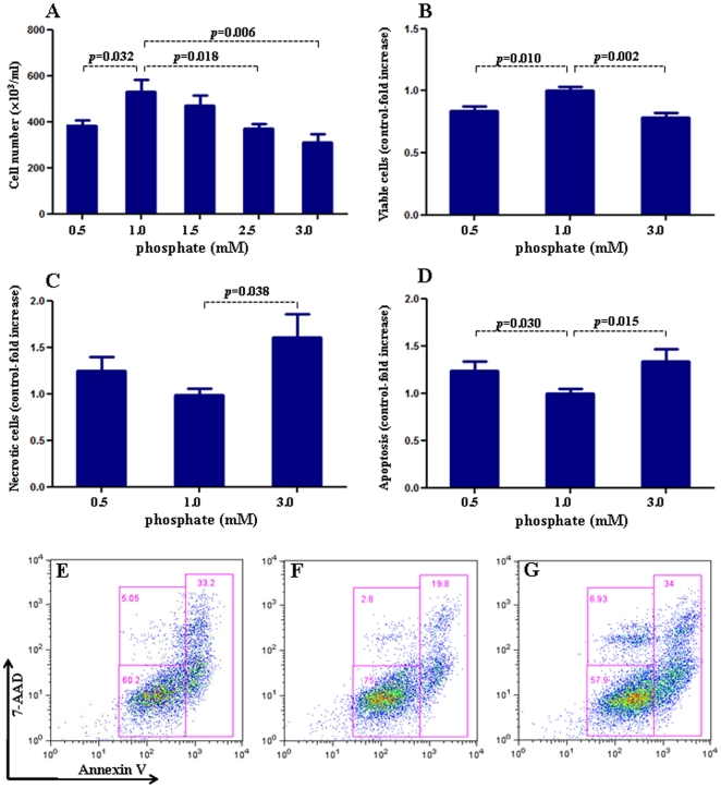
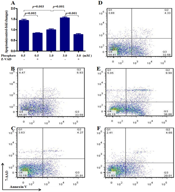
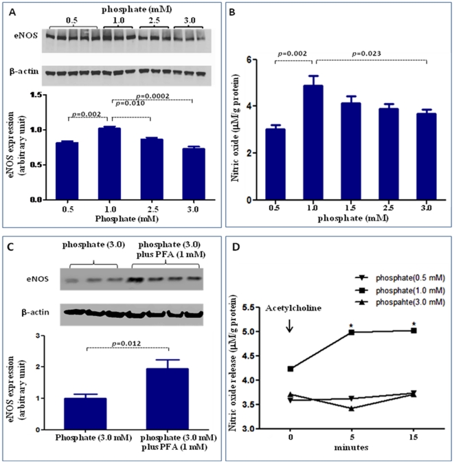
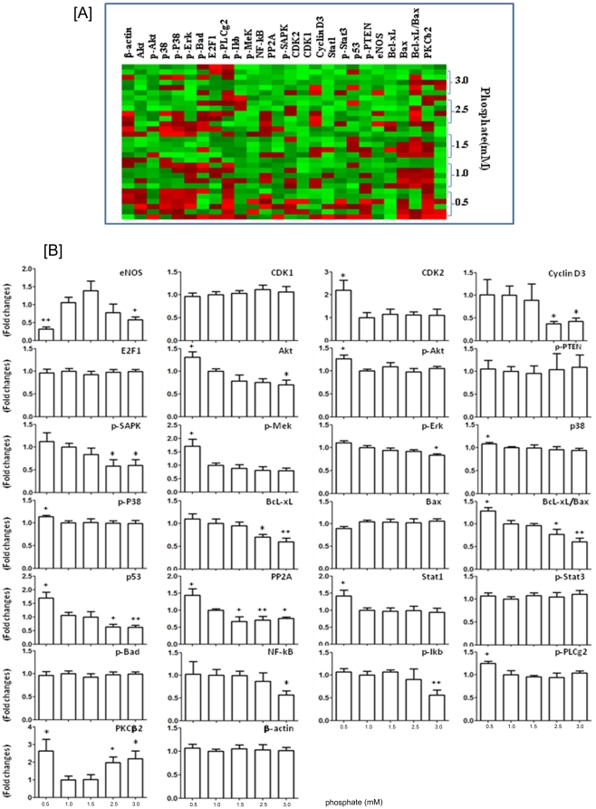
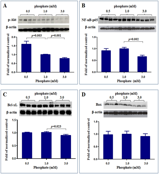
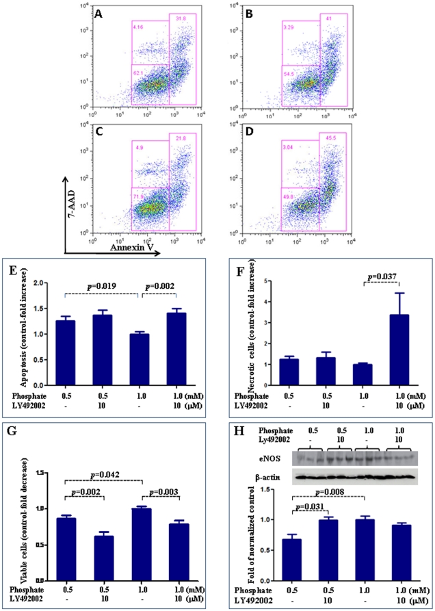
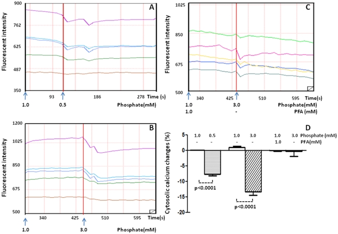
Methodology/principal findings: We studied the effects of simulated hyper- and hypophosphatemia in human umbilical vein endothelial cells in vitro. We found both simulated hyperphosphatemia and hypophosphatemia decrease eNOS expression and NO production. This was associated with reduced intracellular calcium, increased protein kinase C β2 (PKCβ2), reduced cell viability, and increased apoptosis. While simulated hyperphosphatemia was associated with decreased Akt/p-Akt, Bcl-xl/Bax ratios, NFkB-p65 and p-Erk abundance, simulated hypophosphatemia was associated with increased Akt/p-Akt and Bcl-xl/Bax ratios and p-Mek, p38, and p-p38 abundance.

Conclusions/significance: This is the first demonstration of endothelial dysfunction with hypophosphatemia. Our data suggests that both hyperphosphatemia and hypophosphatemia decrease eNOS activity via reduced intracellular calcium and increased PKCβ2. Hyperphosphatemia also appears to reduce eNOS transcription via reduced signaling through PI3K/Akt/NF-kB and MAPK/NF-kB pathways. On the other hand, hypophosphatemia appears to activate these pathways. Our data provides the basis for further studies to elucidate the relationship between altered phosphate homeostasis and cardiovascular disease. As a corollary, our data suggests that the level of phosphate in the culture media, if not in the physiologic range, may inadvertently affect experimental results.


Conflict of interest statement

Competing Interests: The authors have declared that no competing interests exist.

Figures

Figure 1
Figure 1. Effect of inorganic phosphate on endothelial cell proliferation and apoptosis.
We examined the effect of exposure to different concentrations of inorganic phosphate for 24 hours on human umbilical vein endothelial cell proliferation and apoptosis. In Figure 1A, cell number is determined by light microscopy counting of trypan blue-excluding cells, whereas B–D pertain to flow analysis. Incubation in media simulating hypophosphatemia (0.5 mM) or hyperphosphatemia (3 mM phosphate) resulted in a significant reduction of cell numbers (A), decreased cell viability (B) and increased cell dearth (C, statistically significant only seen in hyperphosphatemia) when compared to cells incubated in the medium containing 1 mM phosphate. Similarly, incubation in media simulating hypophosphatemia (0.5 mM, D & E) or hyperphosphatemia (3 mM phosphate, D & G) resulted in a significant increase in apoptotic cells when compared to cells incubated in the medium containing 1 mM phosphate (D & F). In Figure E–G, data in each quadrant are the percentage of total cells, and shown as mean +/− SEM. p values vs control (1 mM phosphate). N = 6∼9.
Figure 2
Figure 2. Effect of pan-caspase inhibitor z-VAD in preventing apoptosis caused by simulated hypophosphatemia and hyperphosphatemia.
To further define the role of inorganic phosphate on human umbilical vein endothelial cell apoptosis, a pan-caspase inhibitor z-VAD (40 uM) was added to the medium containing various concentrations of phosphate. Incubation with pan-caspase inhibitor z-VAD prevented apoptosis induced by both simulated hyperphosphatemia and hypophosphatemia (Figure 2 A–F). B: hypophosphatemia (0.5 mM); C: hypophosphatemia (0.5 mM) plus z-VAD; D: phosphate (1 mM); E: hyperphosphatemia (3 mM phosphate); F: hyperphosphatemia (3 mM) plus z-VAD. In Figure B–F, data in each quadrant are the percentage of total cells, and shown as mean +/− SEM. p values vs control (1 mM phosphate). N = 6∼9.
Figure 3
Figure 3. Effect of inorganic phosphate on eNOS expression and NO production
. We examined the effect of exposure to different concentrations of inorganic phosphate for 24 hours on NO production and eNOS expression. Incubation in media simulating hypophosphatemia (0.5 mM) or hyperphosphatemia (3 mM phosphate) resulted in a significant reduction in eNOS expression (A) and NO production (B). The down-regulation of eNOS following exposure to simulated hyperphosphatemia (3 mM phosphate) was reversed with co-administration of 1 mM PFA, which is a specific inhibitor of phosphate transport across the cell membrane (C). To determine whether abnormal phosphate levels affected the ability of acetylcholine (a physiological activator of eNOS) to stimulate NO production, a set of cells was incubated with 2 µM acetylcholine and the supernatant NO levels before (0 minute) and 5 minute and 15 minute post acetylcholine stimulation was measured. As shown in (D), incubation in medium with 1 mM phosphate resulted in a significant NO production 5 minute post acetylcholine stimulation and remained elevated at 15 minutes. In contrast, incubation in media simulating both hypophosphatemia (0.5 mM) and hyperphosphatemia (3 mM phosphate) completely prevented acetylcholine-induced stimulation of NO generation, pointing to endothelial dysfunction. p values vs control (1 mM phosphate). N = 6∼10.
Figure 4
Figure 4. Effect of inorganic phosphate on normal human lung fibroblast proliferation.
We examined the effect of exposure to different concentrations of inorganic phosphate for 24 hours on normal human lung fibroblast proliferation. The cell proliferation/viability were validated by the mitochondria-dependent reduction of MTT [3-(4,5-dimethylthiazol-2-yl)-2, 5-diphenyl tetrazolium bromide] to purple formazan as detailed in the Method section. Incubation in media simulating hypophosphatemia (0.5 mM) or hyperphosphatemia (3 mM phosphate) had no effect on fibroblast survival when compared to cells incubated in the medium containing 1 mM phosphate.
Figure 5
Figure 5. Effect of inorganic phosphate on cell signaling protein expression.
Using reverse-phase protein microarray, we examined the effect of exposure to different concentrations of inorganic phosphate for 24 hours on human umbilical vein endothelial cell signaling protein expression. (A): shows the reverse-phase protein microarray heat map of key molecules for signaling pathway (green represents expression below median and red represents above-median expression). (B): shows the quantitative signaling protein expression in the endothelial cells incubated for 24 hour with different phosphate concentrations from 0.5 mM to 3.0 mM. Compared to the cells incubated with physiologic phosphate level (1.0 mM), those exposed to simulated hyperphosphatemia (3 mM) showed significantly reduced eNOS, cyclin D3, Akt, p-SAPK, p-Erk, p53, PP2A, NF-kB-p65, p-IkB, Bcl-xl , and Bcl-xl/Bax ratio, but significantly increased level of PKCβ2. On the other hand, the cells exposed to simulated hypophosphatemia (0.5 mM) showed significantly decreased level of eNOS, but significantly increased CDK2, Akt, p-Akt, p-Mek, p38, p-p38, p53, PP2A, Stat1, p-PLCg2, PKCβ2, and Bcl-xl/Bax ratio. *p<0.05, **p<0.01 vs control (1 mM phosphate).
Figure 6
Figure 6. Western blot analysis of p-Akt, NFkB, Bcl-xL and Bax expressions.
To validate the reverse-phase protein microarray data, we examined the protein expression for p-Akt, NF-kB-p65, Bcl-xL and Bax using western blot. Compared to the control cells exposed to physiologic phosphate level (1.0 mM), the cells exposed to simulated hypophospatemia (0.5 mM) showed significantly increased p-Akt (A), while the cells exposed to simulated hyperphospatemia (3 mM) showed significantly reduced p-Akt (A), NF-kB-p65 (B) and Bcl-xL (C). The levels of Bax were not significantly different between the three groups (D). P values vs control (1 mM phosphate). N = 4∼8.
Figure 7
Figure 7. Effect of Akt pathway on endothelial function.
We used PI3K/Akt inhibitor, Ly492002, to explore the role of altered Akt in the endothelial dysfunction induced by simulated hypophosphatemia. Ly492002 had no effect on increased apoptosis induced by low phosphate levels (Figure 5A–E). LY492002 resulted in decreased cell viability in hypophosphatemia. It led to decreased cell viability and increased cell death in physiologic phosphate levels (Figure 5 F & G). On the other hand, Ly492002 was able to block the reduction of eNOS expression induced by simulated hypophospatemia but had no effect on eNOS expression in cells exposed to physiologic phosphate levels (Figure 5H). In Figure A–D, data in each quadrant are the percentage of total cells, and shown as mean +/− SEM. A: 0.5 mM phosphate; B: 1 mM phosphatre; C: 0.5 mM phosphate+Ly492002; D: 1.0 mM phosphate+Ly492002.
Figure 8
Figure 8. Effect of phosphate on intracellular calcium.
The changes of fluorescence intensity was recorded in endothelial cells treated with 0.5 mM phosphate (A), 1 mM phosphate (B) and PFA +3 mM phosphate (C). Percentage changes in intracellular fluorescent intensity is illustrated in D. N = 5 per condition.

References

    1. Farrow EG, White KE. Recent advances in renal phosphate handling. Nat Rev Nephrol. 2010;6:207–217. - PMC - PubMed
    1. Deo R, Fyr CL, Fried LF, Newman AB, Harris TB, et al. Kidney dysfunction and fatal cardiovascular disease–an association independent of atherosclerotic events: results from the Health, Aging, and Body Composition (Health ABC) study. Am Heart J. 2008;155:62–68. - PubMed
    1. Himmelfarb J, Stenvinkel P, Ikizler TA, Hakim RM. The elephant in uremia: oxidant stress as a unifying concept of cardiovascular disease in uremia. Kidney Int. 2002;62:1524–1538. - PubMed
    1. Vaziri ND. Oxidative stress in uremia: nature, mechanisms, and potential consequences. Semin Nephrol. 2004;24:469–473. - PubMed
    1. Vaziri ND. Dyslipidemia of chronic renal failure: the nature, mechanisms, and potential consequences. Am J Physiol Renal Physiol. 2006;290:F262–272. - PubMed

Publication types

MeSH terms