. 2011;6(8):e23282.
doi: 10.1371/journal.pone.0023282. Epub 2011 Aug 16.

Tumor-initiating cells are enriched in CD44(hi) population in murine salivary gland tumor

Affiliations

Tumor-initiating cells are enriched in CD44(hi) population in murine salivary gland tumor

Shukun Shen et al. PLoS One. 2011.

Abstract

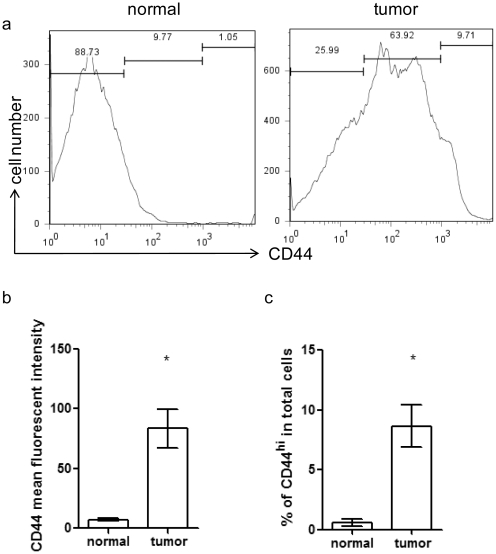
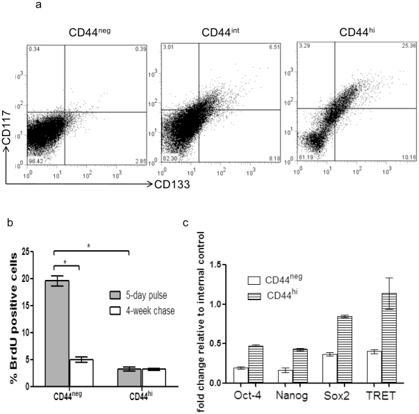
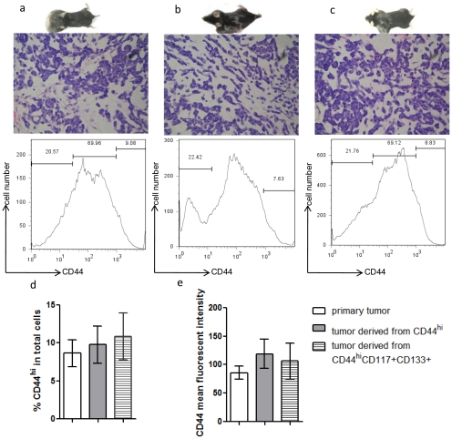
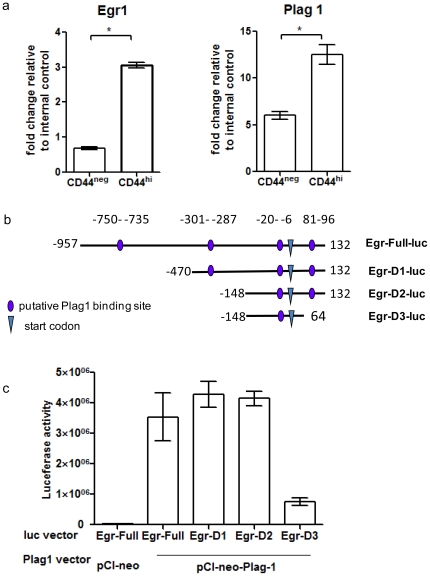
Tumor-initiating cells (T-ICs) discovered in various tumors have been widely reported. However, T-IC populations in salivary gland tumors have yet to be elucidated. Using the established Pleomorphic Adenoma Gene-1 (Plag1) transgenic mouse model of a salivary gland tumor, we identified CD44(high) (CD44(hi)) tumor cells, characterized by high levels of CD44 cell surface expression, as the T-ICs for pleomorphic adenomas. These CD44(hi) tumor cells incorporated 5-bromo-2-deoxyuridine (BrdU), at a lower rate than their CD44(negative) (CD44(neg)) counterparts, and also retained BrdU for a long period of time. Cell surface maker analysis revealed that 25% of the CD44(hi) tumor cells co-express other cancer stem cell markers such as CD133 and CD117. As few as 500 CD44(hi) tumor cells were sufficient to initiate pleomorphic adenomas in one third of the wildtype mice, whereas more than 1×10(4) CD44(neg) cells were needed for the same purpose. In NIH 3T3 cells, Plag1 was capable of activating the gene transcription of Egr1, a known upregulator for CD44. Furthermore, deletion of sequence 81-96 in the Egr1 promoter region abolished the effect of Plag1 on Egr1 upregulation. Our results establish the existence of T-ICs in murine salivary gland tumors, and suggest a potential molecular mechanism for CD44 upregulation.


Conflict of interest statement

Competing Interests: The authors have declared that no competing interests exist.

Figures

Figure 1
Figure 1. Frequency of CD44hi cells in mouse salivary gland tumors.
(a) Histograms of CD44 staining in normal salivary gland cells from wildtype mice (left panel) or tumor salivary gland cells from Plag1 transgenic mice (right panel). Representative graphs from of each group (n = 7) are shown. (b) CD44 mean fluorescent intensity in normal salivary gland cells from wildtype mice (left panel) or tumor salivary gland cells from Plag1 transgenic mice. n = 9, * p<0.05 (normal, vs. tumor, t-test); (c) Percentage of CD44hi cells among total salivary gland cells in normal glands or tumor glands. n = 9, * p<0.05 (normal, vs. tumor, t-test).
Figure 2
Figure 2. Phenotypic analysis of tumor cells.
(a) CD117 and CD133 staining of tumor cells. Mice salivary gland tumor cells were divided into CD44neg, CD44int, and CD44hi subpopulations via FACS; each subpopulation was analyzed for CD117 and CD133 expression. (b) BrdU incorporation. BrdU was injected into tumor-bearing mice at 50 mg/kg twice daily for 5 days, then half of the mice were sacrificed and half were rested for 4 weeks before CD44 and BrdU analysis. (n = 5 each group, * p<0.05 t-test). (c) Comparison of transcript levels for stem cell related genes in CD44neg and CD44hi tumor cells. Transcripts levels for all four genes were higher in CD44hi cells than in CD44neg cells (p<0.05, t-test).
Figure 3
Figure 3. Histological and cell surface marker analysis for secondary tumors.
(a–c) Each panel includes a picture of tumor bearing mouse (top), H&E staining (400×) of the tumor (middle), and tumor cell CD44 staining detected by FACS (bottom). (a) Primary tumors from Plag1 transgenic mice. Mice developed bilateral salivary gland tumors. (b) Tumors derived from CD44hi tumor cell transplant. Mice were injected with 1×10³ CD44hi cells at left side and 1×10³ CD44neg cells at right side as control. Tumors only developed on the left side of the recipients. (c) Tumors derived from CD44hiCD117+CD133+ tumor cells transplant. Mice were injected with 1×10³CD44hiCD117+CD133+ cells at left side and 1×10³ CD44negCD117+CD133+ cells at right side as control. Tumors only developed at left side of the recipients. (d) Percentage of CD44hi cells among total cells in primary tumors (open bar), among total cells in tumors derived from CD44hi cells (grey-color filled bar), and among total cells in tumors derived from CD44hi CD117+CD133+ cells (shaded bar) (n = 9, P>0.05 between any two groups). (e) CD44 mean fluorescent intensity in primary tumors (open bar), in tumors derived from CD44hi cells (grey-color filled bar), and in tumors derived from CD44hi CD117+CD133+ cells (shaded bar) (n = 9, P>0.05 between any two groups).
Figure 4
Figure 4. Plag1 upregulated Egr1 transcription in NIH3T3 cells.
(a) Plag1 and Egr1 transcripts levels in CD44hi and CD44neg tumor cells were determined by real-time PCR. Expression levels were normalized to Ppib gene as internal control (* p<0.05). (b) Schematic diagram of Egr1 promoter region and truncated forms used. Four putative Plag1 binding sequences are marked in the mouse Egr1 promoter region −750 to 132. Egr-Full-luc contains all four sequences, Egr-D1-luc lacked binding sequence in region −750–−735, Egr-D2-luc lacked binding sequence in region −301–−287, Egr-D3-luc lacked binding sequence in region 81–96. (c) Transactivation of Egr1 promoter by Plag1. Co-transfection of vector pCI-neo-Plag1 and luciferase reporter driven by truncated forms of Egr1 promoter in NIH3T3 cells. As expected, pCI-neo empty vector co-transfected with Full-luc did not induce significant luciferase activity. Relative luciferase activity was normalized with β-galactosidase activity derived from pSV-β-galactosidase control vector. Results were shown as average of triplicates.

Similar articles

Cited by

References

    1. Reya T, Morrison SJ, Clarke MF, Weissman IL. Stem cells, cancer, and cancer stem cells. Nature. 2001;414:105–111. - PubMed
    1. Chan KS, Espinosa I, Chao M, Wong D, Ailles L, et al. Identification, molecular characterization, clinical prognosis, and therapeutic targeting of human bladder tumor-initiating cells. Proc Natl Acad Sci U S A. 2009;106:14016–14021. - PMC - PubMed
    1. Dubrovska A, Kim S, Salamone RJ, Walker JR, Maira SM, et al. The role of PTEN/Akt/PI3K signaling in the maintenance and viability of prostate cancer stem-like cell populations. Proc Natl Acad Sci U S A. 2009;106:268–273. - PMC - PubMed
    1. Hong SP, Wen J, Bang S, Park S, Song SY. CD44-positive cells are responsible for gemcitabine resistance in pancreatic cancer cells. Int J Cancer. 2009;125:2323–2331. - PubMed
    1. Bonnet D, Dick JE. Human acute myeloid leukemia is organized as a hierarchy that originates from a primitive hematopoietic cell. Nat Med. 1997;3:730–737. - PubMed

Publication types

MeSH terms