. 2011;6(8):e23351.
doi: 10.1371/journal.pone.0023351. Epub 2011 Aug 16.

A cytoplasmic negative regulator isoform of ATF7 impairs ATF7 and ATF2 phosphorylation and transcriptional activity

Affiliations

A cytoplasmic negative regulator isoform of ATF7 impairs ATF7 and ATF2 phosphorylation and transcriptional activity

Jessica Diring et al. PLoS One. 2011.

Abstract

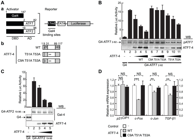
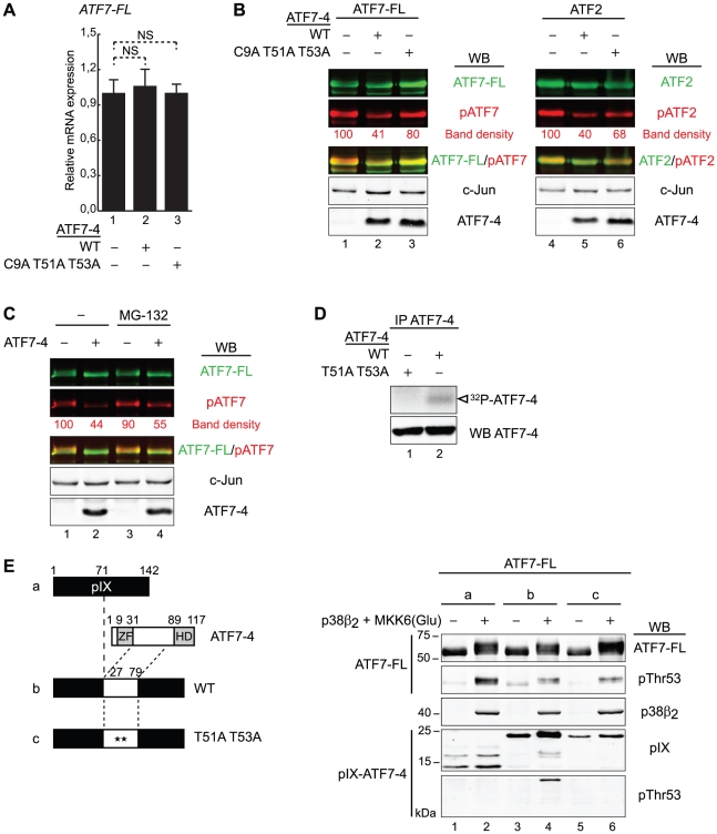
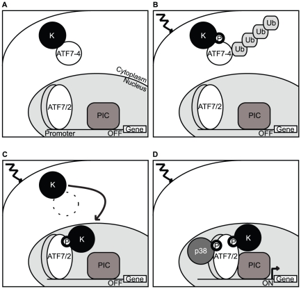
Alternative splicing and post-translational modifications are processes that give rise to the complexity of the proteome. The nuclear ATF7 and ATF2 (activating transcription factor) are structurally homologous leucine zipper transcription factors encoded by distinct genes. Stress and growth factors activate ATF2 and ATF7 mainly via sequential phosphorylation of two conserved threonine residues in their activation domain. Distinct protein kinases, among which mitogen-activated protein kinases (MAPK), phosphorylate ATF2 and ATF7 first on Thr71/Thr53 and next on Thr69/Thr51 residues respectively, resulting in transcriptional activation. Here, we identify and characterize a cytoplasmic alternatively spliced isoform of ATF7. This variant, named ATF7-4, inhibits both ATF2 and ATF7 transcriptional activities by impairing the first phosphorylation event on Thr71/Thr53 residues. ATF7-4 indeed sequesters the Thr53-phosphorylating kinase in the cytoplasm. Upon stimulus-induced phosphorylation, ATF7-4 is poly-ubiquitinated and degraded, enabling the release of the kinase and ATF7/ATF2 activation. Our data therefore conclusively establish that ATF7-4 is an important cytoplasmic negative regulator of ATF7 and ATF2 transcription factors.


Conflict of interest statement

Competing Interests: The authors have declared that no competing interests exist.

Figures

Figure 1
Figure 1. Identification of ATF7-4 as a novel alternatively spliced ATF7 isoform.
(A) The structure of the ATF7 gene, a member of the ATF2/ATF7/CREB5 family, is shown schematically: the twelve exons are indicated by numbered boxes, and an alternative splicing region encompassing exons 4 to 6 is compared between human and mouse species. The sequences of the donor (DS) and acceptor (AS) sites for splicing are indicated, capital and small letters corresponding to the exonic and the intronic parts respectively. 4a and 5a are alternatively included exons adjacent to exons 4 and 5 respectively. The start codon, stop codons and polyadenylation sites are shown. (B) Schematic representation of the exonic structure of three ATF7 alternatively spliced isoforms. The regions encoding the zinc finger (ZnF)/activation domain (AD) or the nuclear localization signal (NLS)/basic region-leucine zipper (b-Zip) are indicated. “+” indicates that the proteins are encoded, either in human or in mouse species. (C) Amino acid sequence alignment of ATF7-4 exon 4 and specific exon 4a of a series of mammalian species. A conserved C-terminal hydrophobic domain is highlighted.
Figure 2
Figure 2. Pattern of expression of ATF7-4 transcript.
(A and B) Real-time RT-PCR analysis of ATF7-4 gene expression profile (left panels) (A) in a 24 human tissue-cDNA array and (B) in a selection of human transformed cell lines. Right panels show the ATF7-4 expression relative to that of ATF7-FL. PBL stands for peripheral blood leucocytes. Data are the average of at least five independent experiments (standard deviations are shown). Data were normalized to β-actin gene expression. (C) Endogenous ATF7-4 protein was analyzed by western-blotting (WB) with a specific anti-ATF7-4 monoclonal antibody in human HeLa cells and mouse 3T3 fibroblasts. HeLa cells were transfected with either the px-ATF7-4 expression vector or a control, or specific siRNA targeting ATF7-4. β-actin was analyzed in parallel as a loading control.
Figure 3
Figure 3. The hydrophobic domain of ATF7-4 controls its cytoplasmic localization.
(A and C) Confocal microscopy images of (A) ATF7-FL, ATF7-4, and (C) eGFP proteins or eGFP-ATF7-4 fusion versions overexpressed in HeLa cells and stained by immunofluorescence with specific antibodies (central panels). DNA was stained with Hoechst 33258 (left panels). A merge of signals is shown on the right panels. (B–D) Analysis of the functional role of the ATF7-4 hydrophobic domain. (B and C) The relocalization of eGFP (lane 1) was assessed for eGFP fusion proteins with ATF7-4 hydrophobic domain (HD), either wild type (wt, lane 2) or mutant version (mt, lane 3). In the latter construct, the four conserved hydrophobic residues (highlighted) were mutated (asterisks) to alanine. Representative confocal images are shown. (D) The fluorescence intensity in the nucleus (N) over the total cell intensity (N+C) was quantified using ImageJ. The results presented are the average of at least 22 cells analyses for each condition. Standard deviations and statistical significance by Student t-test are shown: NS = nonsignificant, ***p<0.001. Scale bar: 10 µm.
Figure 4
Figure 4. ATF7-4 is phosphorylated and subjected to degradation by a proteasome 26S-dependent pathway.
(A) Schematic diagrams indicating the known phosphorylated residues of ATF7-FL protein and the derived series of ATF7-4 mutants used. (B) The indicated WT or mutant versions of ATF7-4 were co-expressed with p38β2 and constitutively active MKK6(Glu) in HeLa cells. Cell lysates were either untreated or treated with calf intestinal phosphatase (CIP), in presence of EDTA where indicated. The analysis was performed by western-blotting (WB) with specific antibodies as specified to detect non-phosphorylated (ATF7-4) and phosphorylated ATF7-4, either on residues Thr51 (pThr51) or Thr53 (pThr53). (C) ATF7-4 (lanes 1–4) and ATF7-FL (lanes 5–8) were co-expressed with p38β2 and MKK6(Glu) in HeLa cells treated with MG-132 proteasome inhibitor as indicated. Cell lysates were analyzed by WB with specific antibodies to detect ATF7-4, ATF7-FL and their phosphorylated forms. (D) ATF7-4 was co-expressed with 6His-Ubiquitin in HeLa cells treated overnight with MG-132. Whole cell extracts (WCE) were purified by a nickel affinity pulldown (Ni-NTA) and analyzed by WB with specific antibodies to detect the poly-ubiquitinated forms of ATF7-4. The asterisks indicate nonspecific signal.
Figure 5
Figure 5. ATF7-4 inhibits the transcriptional activity of ATF7-FL and ATF2.
(A–C) (A) (a) The luciferase activity assays were performed by co-expressing a Gal4-dependent luciferase reporter and Gal4-ATF fusion versions in HeLa cells. Gal4 DNA binding domain (DBD) was fused to the activation domains (AD) of either ATF7 (B) or ATF2 (C). (A) (b) The transcriptional activities were measured in presence of increasing amounts of co-expressed ATF7-4 or mutant versions. (B and C) Cell lysates were assayed for luciferase activity and analyzed in parallel by western-blotting (WB) with specific antibodies. The data of representative experiments are presented (standard deviations are shown). (D) Endogenous ATF7-FL/ATF2 transcriptional activity was measured by a real-time RT-PCR analysis of the expression of known target genes (c-Fos, c-Jun and TGF-β1) or control (p21CIP1). p38β2 and MKK6(Glu) were co-expressed in HeLa cells treated with MG-132. Relative gene expression was assessed in absence (control) or presence of ATF7-4 WT or mutant version. Data were normalized to hypoxanthine-guanine phosphoribosyltransferase (HPRT) gene expression. Results are the average of three independent experiments. Standard deviations and statistical significance by Student t-test are shown: NS = nonsignificant, **p<0.01, ***p<0.001.
Figure 6
Figure 6. ATF7-4 impairs ATF7-FL/ATF2 phosphorylation by retaining a specific kinase.
(A) Endogenous ATF7-FL relative mRNA expression levels were assessed by real-time RT-PCR in HeLa cells, in presence of overexpressed ATF7-4 WT or mutant derivative. Data were normalized to hypoxanthine-guanine phosphoribosyltransferase (HPRT) gene expression. Results are the average of two independent experiments. Standard deviations and statistical significance by Student t-test are shown: NS = nonsignificant. (B and C) The phosphorylation state of overexpressed ATF7-FL and ATF2 was established in presence of ATF7-4 WT or mutant version, in HeLa cells treated (C) or not (B) with MG-132 proteasome inhibitor. The analysis was performed on total cell extracts by western-blotting (WB) with specific antibodies as indicated. Images of total ATF7-FL/ATF2 (green channel) and phosphorylated forms (red channel) were merged with LI-COR Odyssey software. c-Jun levels were used as a loading control. The relative band density of phosphorylated forms was quantified and normalized to c-Jun band density. Results of representative experiments are shown. (D) HeLa cells overexpressing ATF7-4 or mutant version were treated with MG-132. ATF7-4 and the associated proteins were immunoprecipitated with anti-ATF7-4 specific antibody, and assayed for a kinase assay with radiolabelled [γ-32P]ATP. The analysis was performed by autoradiography (top panel) and WB (bottom) in parallel. The white arrow indicates radiolabelled ATF7-4. (E) The phosphorylation state of overexpressed ATF7-FL was analyzed in presence of pIX (a) or pIX-ATF7-4 fusion proteins with the docking site of ATF7-4 for kinases, either WT (b) or mutant version (c). HeLa cells were co-expressed with p38β2 and MKK6(Glu). Cell lysates were analyzed by WB to detect the specific phosphorylation of ATF7-FL (two upper panels) and pIX-ATF7-4 fusion proteins (two lower panels).
Figure 7
Figure 7. Model illustrating the regulatory role of ATF7-4.
(A) Under resting conditions, ATF7-4 retains in the cytoplasm a kinase (K) that is able to phosphorylate ATF7-FL/ATF2 on Thr53/Thr71 respectively, preventing any transcription activity on their target genes. (B) The stimulus-induced phosphorylation of ATF7-4 promotes its poly-ubiquitination and degradation. (C) Our model proposes that the kinase is subsequently released, enters the nucleus and phosphorylates ATF7-FL/ATF2. (D) This first phosphorylation event is necessary for stress-activated p38 recruitment and ATF7-FL/ATF2 phosphorylation on Thr51/Thr69 respectively. The double phosphorylated forms associate efficiently with preinitiation complex (PIC) and are transcriptionally active.

Similar articles

Cited by

References

    1. Hunter T. The age of crosstalk: phosphorylation, ubiquitination, and beyond. Mol Cell. 2007;28:730–738. - PubMed
    1. Hai TW, Liu F, Coukos WJ, Green MR. Distinguishable promoter elements are involved in transcriptional activation by E1a and cyclic AMP. MolCell Biol. 1989;9:4390–4397. - PMC - PubMed
    1. Maekawa T, Sakura H, Kanei-Ishii C, Sudo T, Yoshimura T, et al. Leucine zipper structure of the protein CRE-BP1 binding to the cyclic AMP response element in brain. EMBO J. 1989;8:2023–2028. - PMC - PubMed
    1. Nomura N, Zu YL, Maekawa T, Tabata S, Akiyama T, et al. Isolation and characterization of a novel member of the gene family encoding the cAMP response element-binding protein CRE-BP1. JBiolChem. 1993;268:4259–4266. - PubMed
    1. Gaire M, Chatton B, Kedinger C. Isolation and characterization of two novel, closely related ATF cDNA clones from HeLa cells. Nucleic Acids Res. 1990;18:3467–3473. - PMC - PubMed

Publication types

MeSH terms

LinkOut - more resources