. 2011;6(8):e23360.
doi: 10.1371/journal.pone.0023360. Epub 2011 Aug 17.

A booster vaccine expressing a latency-associated antigen augments BCG induced immunity and confers enhanced protection against tuberculosis

Affiliations

A booster vaccine expressing a latency-associated antigen augments BCG induced immunity and confers enhanced protection against tuberculosis

Bappaditya Dey et al. PLoS One. 2011.

Abstract

Background: In spite of a consistent protection against tuberculosis (TB) in children, Mycobacterium bovis Bacille Calmette-Guerin (BCG) fails to provide adequate protection against the disease in adults as well as against reactivation of latent infections or exogenous reinfections. It has been speculated that failure to generate adequate memory T cell response, elicitation of inadequate immune response against latency-associated antigens and inability to impart long-term immunity against M. tuberculosis infections are some of the key factors responsible for the limited efficiency of BCG in controlling TB.

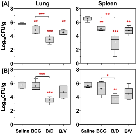
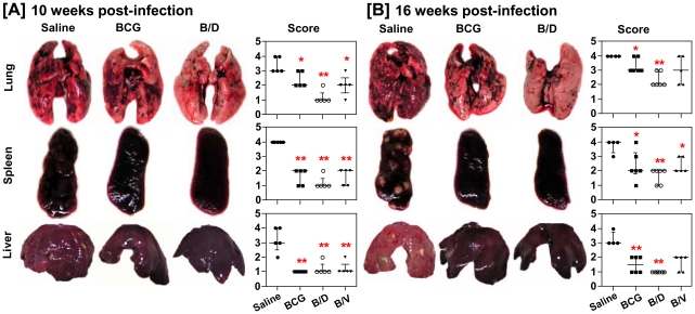
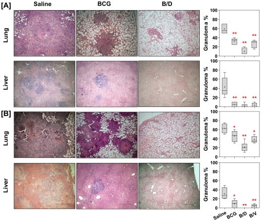
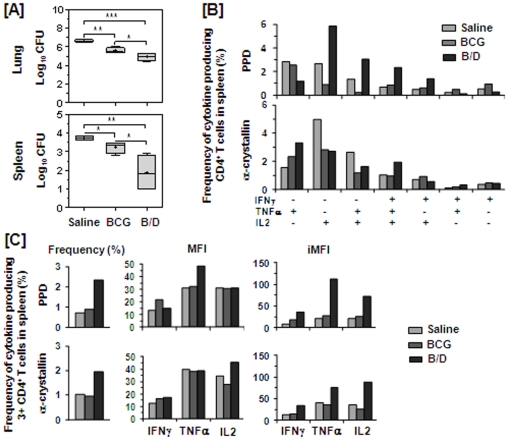
Methods/principal findings: In this study, we evaluated the ability of a DNA vaccine expressing α-crystallin--a key latency antigen of M. tuberculosis to boost the BCG induced immunity. 'BCG prime-DNA boost' regimen (B/D) confers robust protection in guinea pigs along with a reduced pathology in comparison to BCG vaccination (1.37 log(10) and 1.96 log(10) fewer bacilli in lungs and spleen, respectively; p<0.01). In addition, B/D regimen also confers enhanced protection in mice. Further, we show that B/D immunization in mice results in a heightened frequency of PPD and antigen specific multi-functional CD4 T cells (3(+)) simultaneously producing interferon (IFN)γ, tumor necrosis factor (TNF)α and interleukin (IL)2.

Conclusions/significance: These results clearly indicate the superiority of α-crystallin based B/D regimen over BCG. Our study, also demonstrates that protection against TB is predictable by an increased frequency of 3(+) Th1 cells with superior effector functions. We anticipate that this study would significantly contribute towards the development of superior booster vaccines for BCG vaccinated individuals. In addition, this regimen can also be expected to reduce the risk of developing active TB due to reactivation of latent infection.


Conflict of interest statement

Competing Interests: The authors have declared that no competing interests exist.

Figures

Figure 1
Figure 1. Enhanced protection against M. tuberculosis challenge by DNAacr boosting subsequent to BCG vaccination.
For evaluation of protective efficacy two guinea pig experiments were performed. The figure depicts the bacillary load in the lungs and spleen of vaccinated and saline treated guinea pigs at (A) 10 weeks (Exp-I, n = 5) and (B) 16 weeks (Exp-II, n = 6) post-infection. Immunization with B/D regimen resulted in a significantly lower bacillary load in lungs and spleen, when compared to both BCG and saline groups. Log10 CFU were measured and graphically represented by box plot, wherein median values are denoted by horizontal line, the mean is represented by ‘+’, inter quartile range by boxes, and the maximum and minimum values by whiskers. The lower limit of detection was 1.0 log10 CFU/g of tissue and animals with undetectable bacilli were allotted a CFU value of 1.0 log10/g. B/D, BCG prime – DNAacr boost and B/V, BCG prime – vector boost. *, p<0.05, **, p<0.01, ***, p<0.001, when compared to the saline group or as indicated (One-way ANOVA).
Figure 2
Figure 2. Influence of vaccination on gross pathology following M. tuberculosis infection.
The figure depicts representative photographs and graphical depiction of gross scores of lung, liver and spleen of guinea pigs at (A) 10 weeks (n = 5) and (B) 16 weeks (n = 5) post-infection. Based on the extent of involvement, number and size of tubercles, areas of inflammation and necrosis, gross pathological scores were graded from 1 to 4. Each point represents score for an individual animal and the bar depicts median (± inter-quartile range) for each group, *p<0.05 and **p<0.01, when compared to the saline group (Mann-Whitney U test).
Figure 3
Figure 3. Vaccination with B/D regimen reduces granulomatous inflammation in M. tuberculosis infected guinea pigs.
The figure depicts representative photomicrographs (4×) of H&E stained lung and liver sections of guinea pigs at (A) 10 weeks (n = 5) and (B) 16 weeks (n = 5) post-infection. H&E stained lung and liver sections were examined by light microscopy. Lungs: saline group exhibited multi-focal coalescing granulomas with extensive necrosis; BCG group showed discrete granulomas with or without central necrosis and B/D group exhibited negligible and diffused aggregates of inflammatory cells. Liver: saline group showed multiple granulomas, BCG vaccinated animals exhibited mild inflammation and B/D vaccinated animals exhibited no evident signs of inflammation. Scale bar represents 2 mm. Pulmonary and hepatic consolidation were graphically represented as % granuloma by box plot (notations are described in the legend of Fig. 1), *p<0.05 and **p<0.01, when compared to the saline group (Mann-Whitney U test).
Figure 4
Figure 4. Influence of vaccination on the antigen load in pulmonary granulomas post-infection.
The representative photomicrographs (10×) of 5 µm lung sections showing immuno-histochemical staining (brown color) for Ag85 complex proteins in pulmonary granulomas at (A) 10 weeks (Exp-I, n = 5) and (B) 16 weeks (Exp-II, n = 4) post-infection. Unvaccinated animals exhibited extensive staining surrounding and within the granulomatous areas; BCG vaccinated animals exhibited a moderate antigen staining and B/D vaccinated animals exhibited no evident sign of antigen staining in conjunction with negligible granulomatous inflammation. Scale bar represents 1 mm. Extent (Q) of staining was measured [Quick score (Q) = intensity (I) X area (A) of staining] and represented graphically as median (± inter quartile range). *, p<0.05 and **, p<0.01 (Mann-Whitney U test).
Figure 5
Figure 5. Influence of α-crystallin based prime boost vaccination on pulmonary fibrosis following M. tuberculosis infection.
The figure depicts representative photomicrographs (10×) of Van Gieson stained lung sections of guinea pigs euthanized at (A) 10 weeks and (B) 16 weeks post-infection. Saline group exhibited extensive fibrosis or collagen deposition (red color) in granulomatous lesions; BCG group exhibited increased fibrosis with time and B/D group showed complete disease resolution with no evident sign of fibrosis. Scale bar represents 1 mm. Extent (Q) of pulmonary fibrosis was measured by light microscopy [Q = Intensity (I) X area (A) of staining] and represented graphically as median (± inter quartile range). B/D, BCG prime – DNAacr boost. **, p<0.01 (Mann-Whitney U test).
Figure 6
Figure 6. B/D vaccination confers enhanced protection against M. tuberculosis challenge in mice and induces heightened multifunctional CD4 T cell response.
The figure depicts (A) the bacillary load in lungs and spleen of mice (n = 4) at 4 weeks post-infection and (B–C) multifunctional T cell response at 12 weeks post-immunization. Mice experiment was performed once. (A) Log10 CFU is represented by box plot (notations are described in the legend of Fig. 1). The lower limit of detection was 1.0 log10 CFU/g of tissue and animals with undetectable bacilli were allotted a CFU value of 1.0 log10/g. *, p<0.05, **, p<0.01 and ***, p<0.001. (One-way ANOVA). (B–C) T lymphocytes were purified from PPD and α-crystallin stimulated splenocytes (pooled from four mice per group) and stained for CD4 T cell surface marker along with intracellular cytokine staining for IFNγ, TNFα and IL2 followed by FACS analysis. (B) Represents frequency (%) of PPD and α-crystallin specific CD4 T cells expressing each of the seven combinations of IFNγ, TNFα and IL2. (C) Represents frequency of PPD and α-crystallin specific 3+ CD4 T cells along with MFI and iMFI for IFNγ, TNFα and IL2. B/D: BCG prime – DNAacr boost regimen.

Similar articles

Cited by

References

    1. Colditz GA, Berkey CS, Mosteller F, Brewer TF, Wilson ME, et al. The efficacy of bacillus Calmette-Guerin vaccination of newborns and infants in the prevention of tuberculosis: meta-analyses of the published literature. Pediatrics. 1995;96:29–35. - PubMed
    1. Smith PGaMAR., editor. Epidemiology of tuberculosis. Washington, D.C: American Society for Microbiology Press.; 1994. 637
    1. Corbett EL, Watt CJ, Walker N, Maher D, Williams BG, et al. The growing burden of tuberculosis: global trends and interactions with the HIV epidemic. Arch Intern Med. 2003;163:1009–1021. - PubMed
    1. Sherman DR, Voskuil M, Schnappinger D, Liao R, Harrell MI, et al. Regulation of the Mycobacterium tuberculosis hypoxic response gene encoding alpha -crystallin. Proc Natl Acad Sci U S A. 2001;98:7534–7539. - PMC - PubMed
    1. Cunningham AF, Spreadbury CL. Mycobacterial stationary phase induced by low oxygen tension: cell wall thickening and localization of the 16-kilodalton alpha-crystallin homolog. J Bacteriol. 1998;180:801–808. - PMC - PubMed

Publication types

MeSH terms