. 2011;6(8):e23449.
doi: 10.1371/journal.pone.0023449. Epub 2011 Aug 17.

Preterm delivery disrupts the developmental program of the cerebellum

Affiliations

Preterm delivery disrupts the developmental program of the cerebellum

Parthiv Haldipur et al. PLoS One. 2011.

Abstract

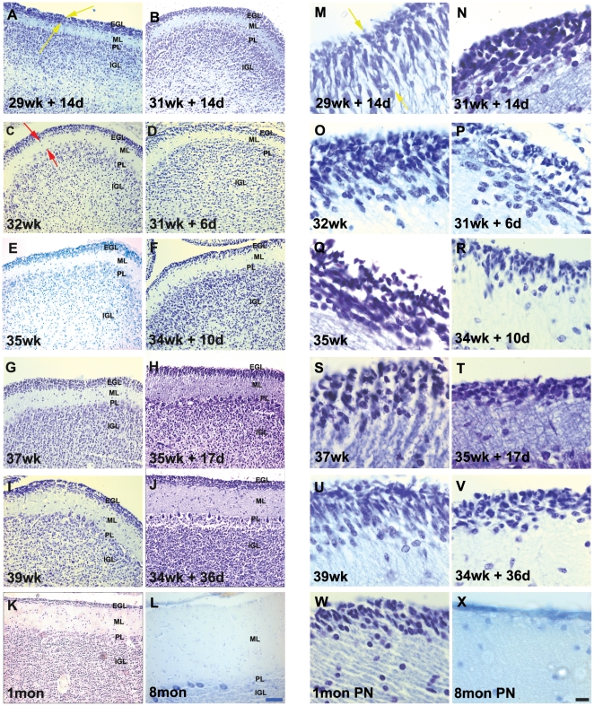
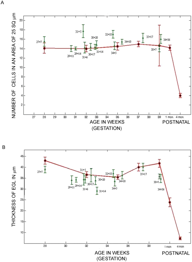
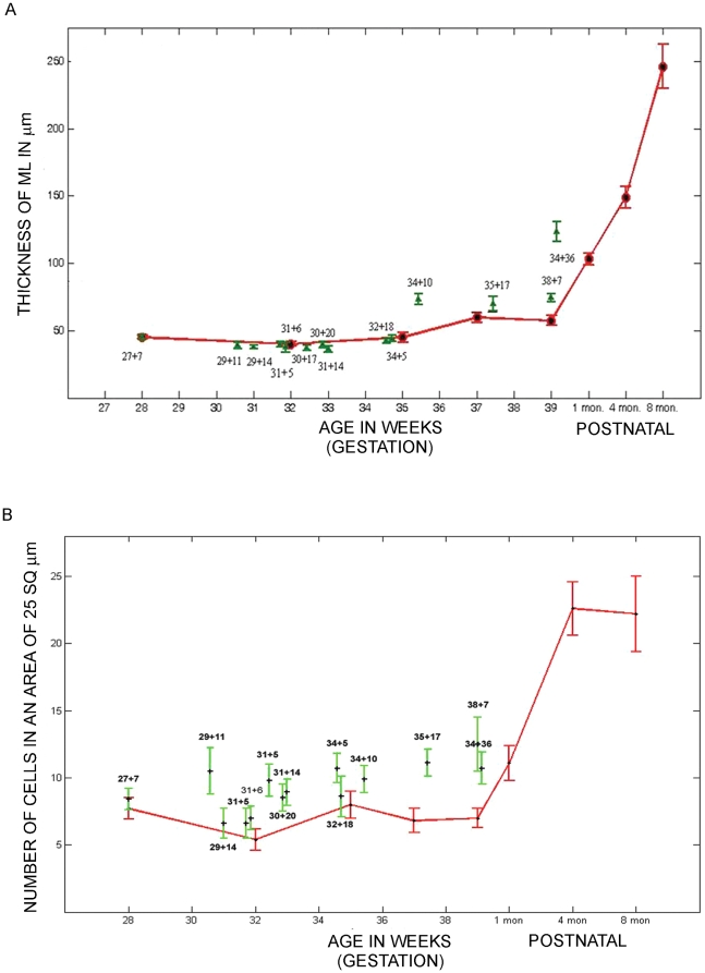
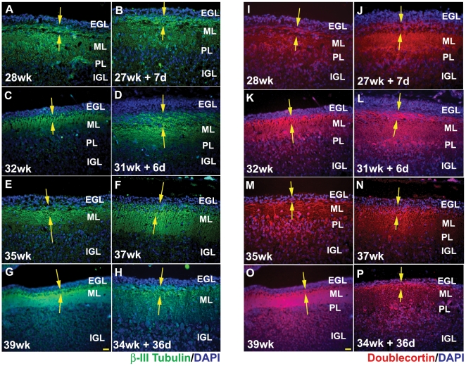
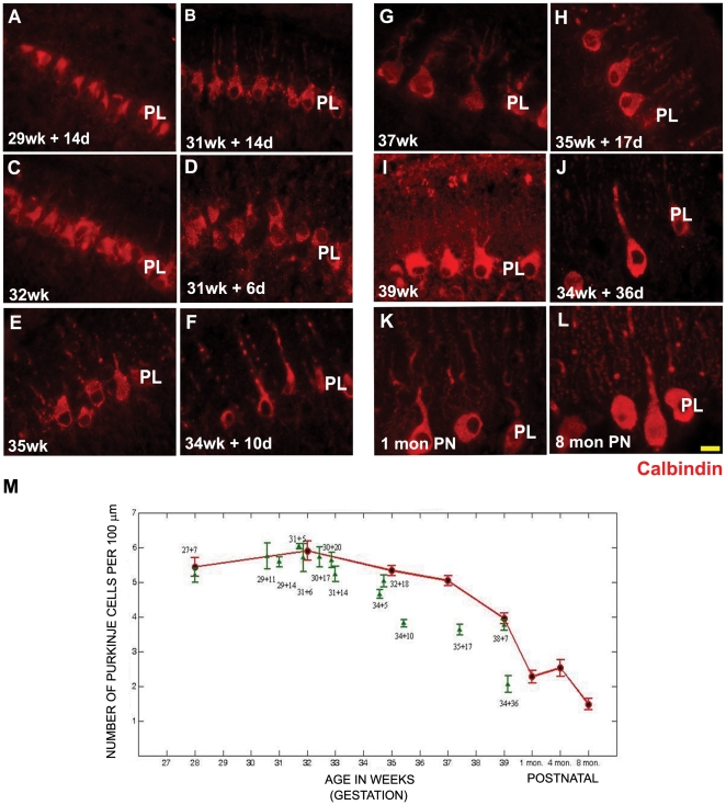
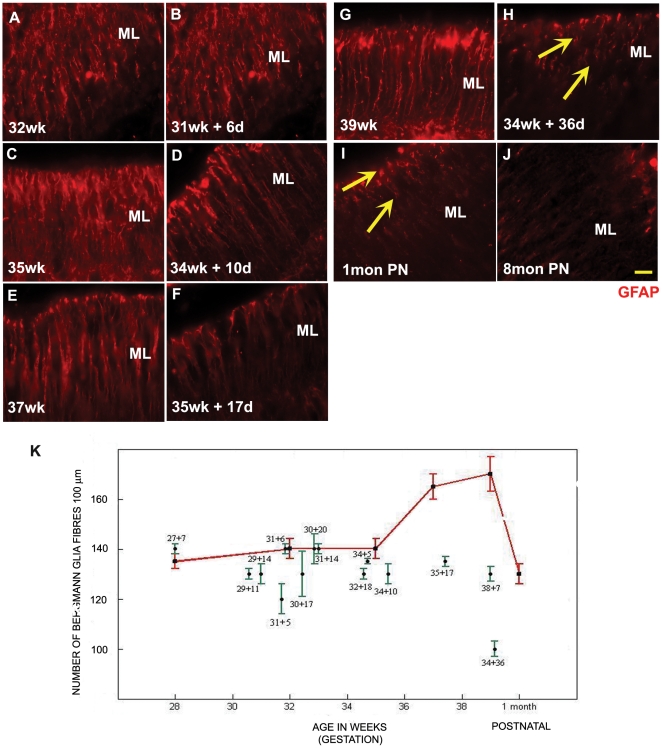
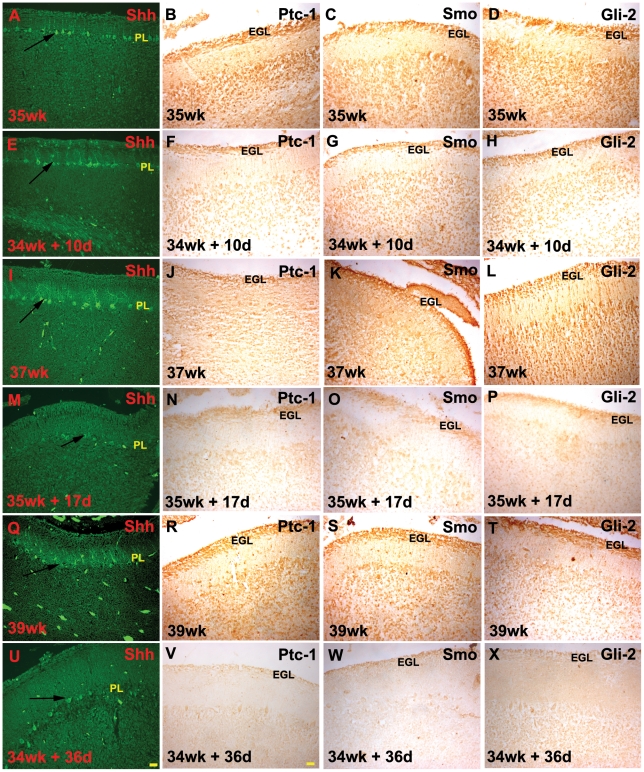
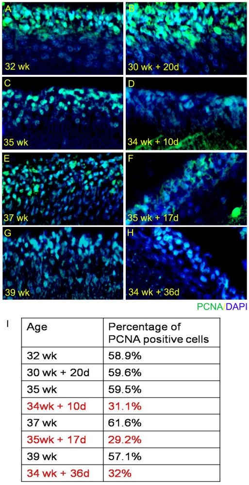
A rapid growth in human cerebellar development occurs in the third trimester, which is impeded by preterm delivery. The goal of this study was to characterize the impact of preterm delivery on the developmental program of the human cerebellum. Still born infants, which meant that all development up to that age had taken place in-utero, were age paired with preterm delivery infants, who had survived in an ex-utero environment, which meant that their development had also taken place outside the uterus. The two groups were assessed on quantitative measures that included molecular markers of granule neuron, purkinje neuron and bergmann glia differentiation, as well as the expression of the sonic hedgehog signaling pathway, that is important for cerebellar growth. We report that premature birth and development in an ex-utero environment leads to a significant decrease in the thickness and an increase in the packing density of the cells within the external granular layer and the inner granular layer well, as a reduction in the density of bergmann glial fibres. In addition, this also leads to a reduced expression of sonic hedgehog in the purkinje layer. We conclude that the developmental program of the cerebellum is specifically modified by events that follow preterm delivery.


Conflict of interest statement

Competing Interests: The authors have declared that no competing interests exist.

Figures

Figure 1
Figure 1. Cresyl violet stained sections of the developing human cerebellum.
(A–X) A greater EGL cell density and reduced EGL thickness were reported in preterms with ex-utero exposure, as compared to their age matched stillborn controls. ML thickness was increased in preterms with ex-utero exposure born after 34 weeks gestation, as compared to their age matched controls. Compare E with F, G with H and I with J. Abbreviations used - wk = number of gestational weeks, d = number of postnatal days, mon PN = postnatal months - born at term. EGL = external granular layer, ML = molecular layer, PL = purkinje cell layer, IGL = internal granular layer. Scale bar = 50 µm (A–L) and 20 µm (M–X).
Figure 2
Figure 2. A significant change in EGL cell density and EGL thickness is seen in samples surviving ex-utero.
(A) Graph showing the density of cells in the EGL versus age (B) Graph showing the thickness of the EGL versus age. Data is shown as mean ± standard deviation of multiple measurements from single sample (SD). Red = still born and infants that were born at term and survived postnatally (1–8 months). Green = preterms that survived in an ex-utero environment.
Figure 3
Figure 3. IGL cell density is increased, although there is no significant change in ML thickness in the samples surviving ex-utero.
(A) Graph showing the thickness of the ML versus age. (B) Graph showing the density of cells in the IGL versus age. Data is shown as mean ± standard deviation of multiple measurements from single sample (SD). Red = still born and infants that were born at term and survived postnatally (1–8 months). Green = preterms that survived in an ex-utero environment.
Figure 4
Figure 4. Ex-utero environment does not alter the expression of EGL differentiation markers β-III tubulin and doublecortin. β-III Tubulin (Green) (A–H) and doublecortin (Red) (I–P) positive cells in the inner EGL (yellow arrows).
There is no drastic difference seen in the number of β-III Tubulin and doublecortin positive cells in the EGL of stillborn controls and preterms that have survived in an ex-utero environment. DAPI staining was used to get a measure of the total number of cells in the EGL. Blue = DAPI. Abbreviations used - wk = number of gestational weeks, d = number of postnatal days. EGL = external granular layer, ML = molecular layer, PL = purkinje cell layer, IGL = internal granular layer. Scale bar = 50 µm.
Figure 5
Figure 5. Calbindin positive purkinje cells of the cerebellum.
There is no statistically significant difference in the number of calbindin positive purkinje cells of the cerebellum of preterms with ex-utero survival in comparison to their age matched still born controls (A–L). However the purkinje cell number was seen to decrease in preterms with ex-utero exposure, born after 34 weeks gestation, as compared to their age matched controls. The sections have been stained using calbindin antibody raised in rabbit (red). Abbreviations used - wk = number of gestational weeks, d = number of postnatal days, mon PN = postnatal months - born at term PL = purkinje cell layer. Scale bar = 20 µm (M) Graph showing the number of calbindin positive cells in the PL of the human cerebellum versus age. Data is shown as mean ± standard deviation of multiple measurements from single sample (SD). Red = still born and postnatal survival born at term. Green = preterms that survived in an ex-utero environment.
Figure 6
Figure 6. Bergmann glia fibre count is reduced in samples surviving ex-utero.
The number of GFAP positive bergmann glia fibres in the ML of the human cerebellum (A–J), was found to reduce in preterms with ex-utero survival in comparison to their age matched still born controls. The morphology of the fibres from the individual with maximum ex-utero exposure (34 g.w+36 d) was found to be similar to those seen in a 1 month postnatal cerebellum in an individual born at term; Compare H with I, arrows. Abbreviations used - wk = number of gestational weeks, d = number of postnatal days, PN = postnatal months- born at term, PL = purkinje cell layer. The sections have been stained using GFAP antibody raised in rabbit (red). Scale bar = 20 µm (K) Graph showing the number of GFAP positive bergmann glia fibres in the ML of the human cerebellum versus age. Data is shown as mean ± standard deviation of multiple measurements from single sample (SD). Red = still born and postnatal survival born at term. Green = preterms that survived in an ex-utero environment.
Figure 7
Figure 7. Distribution of sonic hedgehog (Shh), patched-1 (Ptc), smoothened (Smo) and Gli-2 across ages.
The levels of Shh, its receptor Ptc and its downstream effectors were seen to decrease in preterms with maximum ex-utero exposure, in comparison to their age matched controls. Compare 37 wk (I–L) with 35 wk+17 d (M–P) and 39 wk (Q–T) with 34 wk+36 d (U–X); Abbreviations used - wk = number of gestational weeks, d = number of postnatal days. EGL = external granular layer, ml = molecular layer, PL = purkinje cell layer, IGL = internal granular layer. Sections were stained by immunofluorescence for Shh, while Ptc, Smo and Gli-2 reactivity were developed using the peroxidase method. Scale bar = 50 µm.
Figure 8
Figure 8. The number of PCNA positive cells decreases with reduction in Shh signalling.
While we find that there is no major difference in the number of proliferating population between (A) 32 weeks and (B) 30 wk+20 d, there is a significant difference in the preterms with reduced Shh signalling. For example, compare (C) 35 wk with (D) 34 wk+10 d; (E) 37 wk with (F) 35 wk+17 d; and (G) 39 wk with (H) 34 wk+36 d. Sections were stained by immunofluorescence for PCNA (green) and mounted using vectashield DAPI (blue).

References

    1. Ben-Yehudah G, Fiez JA. Impact of cerebellar lesions on reading and phonological processing. Ann N Y Acad Sci. 2008;1145:260–274. - PubMed
    1. Bugalho P, Correa B, Viana-Baptista M. Role of the cerebellum in cognitive and behavioural control: scientific basis and investigation models. Acta Med Port. 2006;19:257–267. - PubMed
    1. Miall RC, Reckess GZ, Imamizu H. The cerebellum coordinates eye and hand tracking movements. Nat Neurosci. 2001;4:638–644. - PubMed
    1. Cox C, Teknos TN, Barrios M, Brewer GJ, Dick RD, et al. The role of copper suppression as an antiangiogenic strategy in head and neck squamous cell carcinoma. Laryngoscope. 2001;111:696–701. - PubMed
    1. Gordon N. The cerebellum and cognition. Eur J Paediatr Neurol. 2007;11:232–234. - PubMed

Publication types