. 2011;6(8):e23479.
doi: 10.1371/journal.pone.0023479. Epub 2011 Aug 9.

The RelA/SpoT homolog (RSH) superfamily: distribution and functional evolution of ppGpp synthetases and hydrolases across the tree of life

Affiliations

The RelA/SpoT homolog (RSH) superfamily: distribution and functional evolution of ppGpp synthetases and hydrolases across the tree of life

Gemma C Atkinson et al. PLoS One. 2011.

Abstract

RelA/SpoT Homologue (RSH) proteins, named for their sequence similarity to the RelA and SpoT enzymes of Escherichia coli, comprise a superfamily of enzymes that synthesize and/or hydrolyze the alarmone ppGpp, activator of the "stringent" response and regulator of cellular metabolism. The classical "long" RSHs Rel, RelA and SpoT with the ppGpp hydrolase, synthetase, TGS and ACT domain architecture have been found across diverse bacteria and plant chloroplasts, while dedicated single domain ppGpp-synthesizing and -hydrolyzing RSHs have also been discovered in disparate bacteria and animals respectively. However, there is considerable confusion in terms of nomenclature and no comprehensive phylogenetic and sequence analyses have previously been carried out to classify RSHs on a genomic scale. We have performed high-throughput sensitive sequence searching of over 1000 genomes from across the tree of life, in combination with phylogenetic analyses to consolidate previous ad hoc identification of diverse RSHs in different organisms and provide a much-needed unifying terminology for the field. We classify RSHs into 30 subgroups comprising three groups: long RSHs, small alarmone synthetases (SASs), and small alarmone hydrolases (SAHs). Members of nineteen previously unidentified RSH subgroups can now be studied experimentally, including previously unknown RSHs in archaea, expanding the "stringent response" to this domain of life. We have analyzed possible combinations of RSH proteins and their domains in bacterial genomes and compared RSH content with available RSH knock-out data for various organisms to determine the rules of combining RSHs. Through comparative sequence analysis of long and small RSHs, we find exposed sites limited in conservation to the long RSHs that we propose are involved in transmitting regulatory signals. Such signals may be transmitted via NTD to CTD intra-molecular interactions, or inter-molecular interactions either among individual RSH molecules or among long RSHs and other binding partners such as the ribosome.


Conflict of interest statement

Competing Interests: The authors have declared that no competing interests exist.

Figures

Figure 1
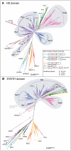
Figure 1. Maximum likelihood phylogenies of the ppGpp synthetase and hydrolase domains.
Trees were generated from RaxML analyses of alignments of A) the ppGpp hydrolase (HD) domain-containing dataset (168 amino acid positions, 1535 sequences), and B) the ppGpp synthetase (SYNTH) domain-containing dataset (670 amino acid positions, 1706 sequences). In both trees, subgroups are labeled and shading behind the branches shows the most common domain structure observed for those groups, as per the legend in the inset box. Symbols on branches indicate bootstrap support, as per the inset box. In cases where the whole subgroup carries both the HD and SYNTH domain (Rel, SpoT, RelA, Rsh1-4, RshA-D), bootstrap support comes from the full length long RSH tree (supplementary file SI2). Branch length is proportional to the number of substitutions per site (see scale bar).
Figure 2
Figure 2. Consensus alignment of long RSHs, with RelSeq NTD structure colored according to conservation patterns in the alignment.
A) Domain structure of the long RSHs, with domain lengths to scale with S. equisimilis Rel[HS]. B) Alignment of long RSH sequences at the 70% level. Secondary structure is shown below the alignment, with “)” characters indicating helices and “>” characters indicating sheets. Secondary structure is obtained from the structure of Rel until position 362, after which second Psipred was used to predict the secondary structure. Disordered regions in the structure are underlaid with a pale grey box, and disordered regions predicted with Disopred are a darker grey. Highlighting of residue columns indicates conservation patterns (also see inset box). Blue highlighting indicates sites that are conserved across all long RSHs. Green highlighting shows those sites that are distinctive in RelA[hS] (strongly differentially conserved or conserved only in RelA[hS]). Yellow highlighted sites are well conserved in Rel[HS]+SpoT[HS] but less so RelA[hS], while purple highlighted sites are well conserved in Rel[HS]+RelA[hS] and less so in SpoT[HS]. Lines beneath the alignment indicate domains with the following colours: dark blue – HD, red – SYNTH, light blue – TGS, green – helical, turquoise – CC, magenta – ACT. Blue and red boxes show sites of the HD and SYNTH nucleotide binding pockets, respectively. Colored boxes in the TGS and ACT domain surround the usually most conserved blocks of these domains as per sequence logos in the Pfam database. The turquoise box in the CC domain indicates the most conserved block of this domain, which also contains the conserved cysteines of . The orange bar above the alignment shows the location of the differentially conserved motif of . Black boxes around RelA[hS] and SpoT[HS] residues show sites that have experienced shifts in substitution rate, as predicted with Diverge. C) Structure of the Rel[HS] protein from S. equisimilis (RelSeq) , colored according to the conservation patterns of the alignment in B.
Figure 3
Figure 3. Schematic diagram for the evolution of long RSHs in bacteria.
Thick gray branches indicate the divergence of bacterial groups, while the inner line shows the divergence of long RSH proteins and their functionality, as per the inset box.
Figure 4
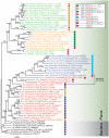
Figure 4. Bayesian inference phylogeny of plant RSHs.
The tree was generated from a MrBayes analysis of 470 amino acid positions from 66 sequences. Colored sequence names indicate subgroups as follows: red – Rsh1, green – Rsh2, orange – Rsh3, blue – Rsh4, and black – bacterial Rel. Numbers on branches show support in the following format: BIPP/MLBP. Support is only shown for branches with BIPP>0.8. Branch length is proportional to the number of substitutions per site (see scale bar).
Figure 5
Figure 5. Bayesian inference phylogeny of the SAHs Mesh1 and Mesh1L.
The tree was generated from a MrBayes analysis of 179 amino acid positions from 99 sequences. Branch support and length are shown as described in Fig. 4. Sequence names are colored by taxonomic groups.
Figure 6
Figure 6. Consensus alignment of long RSH and small RSH subgroups across the ppGpp synthetase and hydrolase domains, with RelSeq NTD structure colored according to conservation patterns in the alignment.
A) Alignment of RSH NTD sequences at the 70% level. Yellow highlighting shows those residues that are only conserved only in long RSHs. Blue and red boxes show sites of the HD and SYNTH nucleotide binding pockets, respectively. Bright turquoise and orange boxes show the location of surface residues in the SYNTH and HD domains respectively that are likely to be involved in inter molecular interactions, or interactions with the CTD in long RSHs. The box is dotted where the region is disordered in the structure. The pale marine box shows those regions that appear to be involved in HD-SYNTH interactions. Arrows show especially interesting sites (see inset box in B). The orange bar above the alignment shows the location of the differentially conserved motif of . B) Structure of the Rel[HS] protein from S. equisimilis (RelSeq) , colored according to the conservation patterns of the alignment in A. The inset box shows a subset of particularly interesting sites (labeled with arrows in A). Residue numbering is as in RelSeq, followed by alignment coordinates from A in parentheses.

References

    1. Pesavento C, Hengge R. Bacterial nucleotide-based second messengers. Curr Opin Microbiol. 2009;12:170–176. - PubMed
    1. Gomelsky M. cAMP, c-di-GMP, c-di-AMP and now cGMP: bacteria use them all! Mol Microbiol. 2011;79:562–565. - PMC - PubMed
    1. Mittenhuber G. Comparative genomics and evolution of genes encoding bacterial (p)ppGpp synthetases/hydrolases (the Rel, RelA and SpoT proteins). J Mol Microbiol Biotechnol. 2001;3:585–600. - PubMed
    1. Gallant J, Palmer L, Pao CC. Anomalous synthesis of ppGpp in growing cells. Cell. 1977;11:181–185. - PubMed
    1. English BP, Hauryliuk V, Sanamrad A, Tankov S, Dekker NH, et al. Proc Natl Acad Sci U S A; 2011. Single-molecule investigations of the stringent response machinery in living bacterial cells. - PMC - PubMed

Publication types

MeSH terms

Substances

LinkOut - more resources