. 2011;6(8):e23524.
doi: 10.1371/journal.pone.0023524. Epub 2011 Aug 15.

Vitamin D receptor deficiency enhances Wnt/β-catenin signaling and tumor burden in colon cancer

Affiliations

Vitamin D receptor deficiency enhances Wnt/β-catenin signaling and tumor burden in colon cancer

María Jesús Larriba et al. PLoS One. 2011.

Abstract

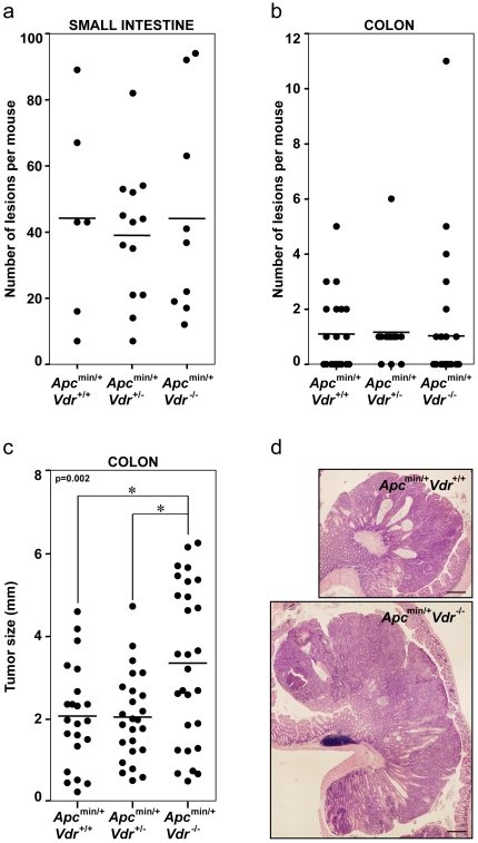
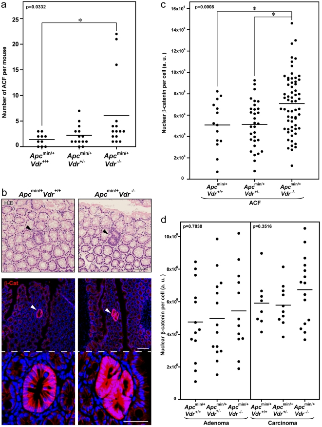
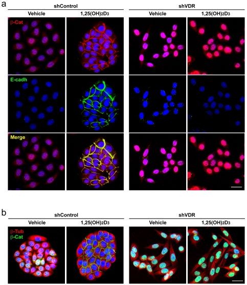
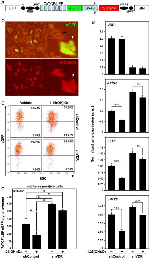
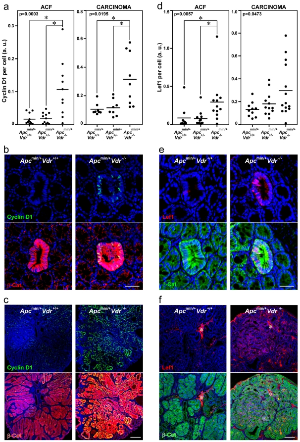
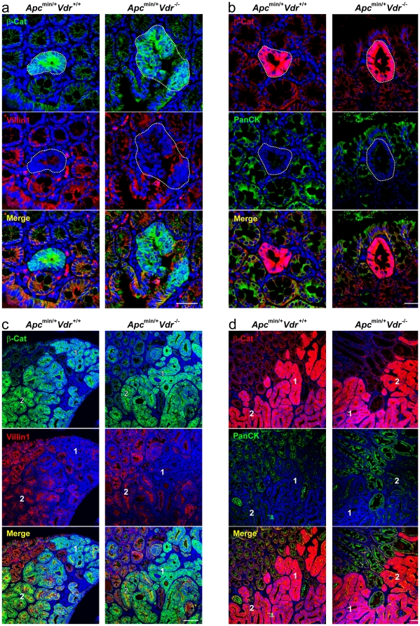
Aberrant activation of the Wnt/β-catenin pathway is critical for the initiation and progression of most colon cancers. This activation provokes the accumulation of nuclear β-catenin and the induction of its target genes. Apc(min/+) mice are the most commonly used model for colon cancer. They harbor a mutated Apc allele and develop intestinal adenomas and carcinomas during the first months of life. This phenotype is caused by the mutation of the second Apc allele and the consequent accumulation of nuclear β-catenin in the affected cells. Here we describe that vitamin D receptor (VDR) is a crucial modulator of nuclear β-catenin levels in colon cancer in vivo. By appropriate breeding of Apc(min/+) mice and Vdr(+/-) mice we have generated animals expressing a mutated Apc allele and two, one, or none Vdr wild type alleles. Lack of Vdr increased the number of colonic Aberrant Crypt Foci (ACF) but not that of adenomas or carcinomas in either small intestine or colon. Importantly, colon ACF and tumors of Apc(min/+)Vdr(-/-) mice had increased nuclear β-catenin and the tumors reached a larger size than those of Apc(min/+)Vdr(+/+). Both ACF and carcinomas in Apc(min/+)Vdr(-/-) mice showed higher expression of β-catenin/TCF target genes. In line with this, VDR knock-down in cultured human colon cancer cells enhanced β-catenin nuclear content and target gene expression. Consistently, VDR depletion abrogated the capacity of 1,25(OH)(2)D(3) to promote the relocation of β-catenin from the nucleus to the plasma membrane and to inhibit β-catenin/TCF target genes. In conclusion, VDR controls the level of nuclear β-catenin in colon cancer cells and can therefore attenuate the impact of oncogenic mutations that activate the Wnt/β-catenin pathway.


Conflict of interest statement

Competing Interests: The authors have declared that no competing interests exist.

Figures

Figure 1
Figure 1. Vdr deficiency increases tumor load but not tumor number in Apcmin/+ mice.
Total number of tumors (adenomas and carcinomas) in the small intestine (a) or in the colon (b) of Apcmin/+Vdr+/+, Apcmin/+Vdr+/− and Apcmin/+Vdr-/- mice. Each dot corresponds to one mouse analyzed and the horizontal line indicates the mean. (c) Size of colon tumors (adenomas and carcinomas) from Apcmin/+Vdr+/+, Apcmin/+Vdr+/- and Apcmin/+Vdr-/- mice. Each dot corresponds to one tumor analyzed and the horizontal line indicates the mean. The p-value for one-way ANOVA analysis is shown. Asterisks indicate significant differences between groups upon Bonferroni multiple comparison post-test. (d) Representative hematoxylin/eosin staining images of colon carcinomas from Apcmin/+Vdr+/+ and Apcmin/+Vdr-/- mice. Scale bar, 300 µm.
Figure 2
Figure 2. β-Catenin nuclear level is elevated in colonic lesions of Apcmin/+Vdr-/- mice.
(a) Total number of colonic ACF in Apcmin/+Vdr+/+, Apcmin/+Vdr+/− and Apcmin/+Vdr-/- mice. Each dot corresponds to one mouse analyzed and the horizontal line indicates the mean. (b) Upper panels, representative hematoxylin/eosin staining images of colonic ACF in Apcmin/+Vdr+/+ and Apcmin/+Vdr-/- mice. Arrowheads indicate the ACF. Scale bar, 50 µm. Middle panels, representative immunofluorescence images showing β-catenin expression and localization in colonic ACF from Apcmin/+Vdr+/+ and Apcmin/+Vdr-/- mice. Arrowheads indicate the ACF. Scale bar, 100 µm. Lower panels are a magnification of middle panels. Scale bar, 50 µm. (c) Quantification of β-catenin nuclear level in colonic ACF from Apcmin/+Vdr+/+, Apcmin/+Vdr+/- and Apcmin/+Vdr-/- mice. Each dot corresponds to one lesion analyzed and the horizontal line indicates the mean. (d) Quantification of β-catenin nuclear level in colonic adenomas and carcinomas from Apcmin/+Vdr+/+, Apcmin/+Vdr+/- and Apcmin/+Vdr-/- mice. Each dot corresponds to one lesion analyzed and the horizontal line indicates the mean. (a, c, d) The p-values for one-way ANOVA analysis are shown. Asterisks indicate significant differences between groups upon Bonferroni multiple comparison post-test.
Figure 3
Figure 3. VDR knock-down in SW480-ADH human colon cancer cells increases β-catenin nuclear level.
Representative immunofluorescence images showing β-catenin and E-cadherin (a) or β-catenin and β-tubulin (b) expression and localization in SW480-ADH cells infected with lentiviruses expressing shVDR or shControl and treated with 1,25(OH)2D3 or vehicle for 72 h. Scale bar, 20 µm.
Figure 4
Figure 4. VDR knock-down in SW480-ADH cells enhances β-catenin/TCF transcriptional activity and reduces the inhibitory effect of 1,25(OH)2D3 on Wnt/β-catenin pathway.
(a) Diagram of the 7xTCF/LEF-eGFP-SV40-mCherry lentiviral construct used to infect shControl and shVDR SW480-ADH cells. (b) Representative phase-contrast and fluorescence images showing the expression of eGFP and mCherry in SW480-ADH cells infected with the 7xTCF/LEF-eGFP-SV40-mCherry vector. Arrowheads indicate similarly infected cells that have variable expression of eGFP. Scale bars, 20 µm. (c) Flow cytometry analysis of eGFP expression in shControl and shVDR SW480-ADH cells infected (mCherry positive) with the 7xTCF/LEF-eGFP-SV40-mCherry vector and treated with 1,25(OH)2D3 or vehicle for 96 h. Percentage of cells in each gate is indicated. (d) Quantification of the flow cytometry data showing the average expression of eGFP in the infected mCherry-positive cells. Data correspond to three independent experiments. The p-value for one-way ANOVA analysis is shown. Asterisks indicate significant differences between groups upon Bonferroni multiple comparison post-test. (e) Analysis by qRT-PCR of VDR, AXIN2, LEF1 and c-MYC mRNA expression in shControl and shVDR SW480-ADH cells treated with 1,25(OH)2D3 or vehicle for 48 h. The geometric average of the expression of SDHA and TBP housekeeping genes was used for RNA expression normalization as indicated in Materials and Methods. Numbers indicate the percentage of inhibition by 1,25(OH)2D3 treatment in each cell type. Data correspond to three independent experiments.
Figure 5
Figure 5. Colonic ACF and carcinomas in Apcmin/+Vdr-/- mice have elevated expression of β-catenin/TCF target genes Ccnd1/Cyclin D1 and Lef1.
Quantification of Cyclin D1 (a) and Lef1 (d) protein expression in colonic lesions from Apcmin/+Vdr+/+, Apcmin/+Vdr+/- and Apcmin/+Vdr-/- mice. Each dot corresponds to one lesion analyzed and the horizontal line indicates the mean. The p-values for one-way ANOVA analysis are shown. Asterisks indicate significant differences between groups upon Bonferroni multiple comparison post-test. Representative immunofluorescence images showing β-catenin and Cyclin D1 (b, c) or β-catenin and Lef1 (e, f) expression and localization in colon ACF (b, e) and carcinomas (c, f) from Apcmin/+Vdr+/+ and Apcmin/+Vdr-/- mice. Scale bars, 50 µm (b, e) and 200 µm (c, f). Asterisks indicate an unspecific staining.
Figure 6
Figure 6. Vdr depletion does not affect the differentiation status of colon ACF and carcinomas in Apcmin/+ mice.
Representative immunofluorescence images showing β-catenin and villin1 (a, c) or β-catenin and pan-cytokeratin (b, d) expression and localization in colon ACF (a, b) and carcinomas (c, d) from Apcmin/+Vdr+/+ and Apcmin/+Vdr-/- mice. Dotted-lines delimit ACF. Numbers indicate regions with different pattern of expression of the analyzed proteins within the same tumor: 1) high β-catenin, low villin1/cytokeratins; 2) low β-catenin, high villin1/cytokeratins. Scale bars, 50 µm (a, b) and 200 µm (c, d).

References

    1. Jemal A, Bray F, Center MM, Ferlay J, Ward E, et al. Global cancer statistics. CA Cancer J Clin. 2011;61:69–90. - PubMed
    1. Fodde R, Smits R, Clevers H. APC, signal transduction and genetic instability in colorectal cancer. Nat Rev Cancer. 2001;1:55–67. - PubMed
    1. Humphries A, Wright NA. Colonic crypt organization and tumorigenesis. Nat Rev Cancer. 2008;8:415–424. - PubMed
    1. Su LK, Kinzler KW, Vogelstein B, Preisinger AC, Moser AR, et al. Multiple intestinal neoplasia caused by a mutation in the murine homolog of the APC gene. Science. 1992;256:668–670. - PubMed
    1. Gregorieff A, Clevers H. Wnt signaling in the intestinal epithelium: from endoderm to cancer. Genes Dev. 2005;19:877–890. - PubMed

Publication types

MeSH terms