. 2011;6(8):e23535.
doi: 10.1371/journal.pone.0023535. Epub 2011 Aug 12.

A gain-of-function germline mutation in Drosophila ras1 affects apoptosis and cell fate during development

Affiliations

A gain-of-function germline mutation in Drosophila ras1 affects apoptosis and cell fate during development

Christopher Gafuik et al. PLoS One. 2011.

Abstract

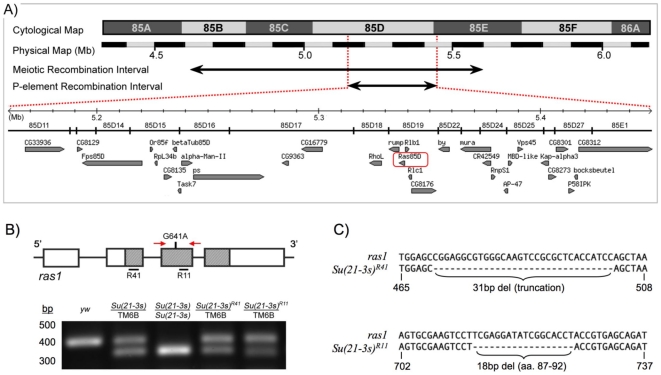
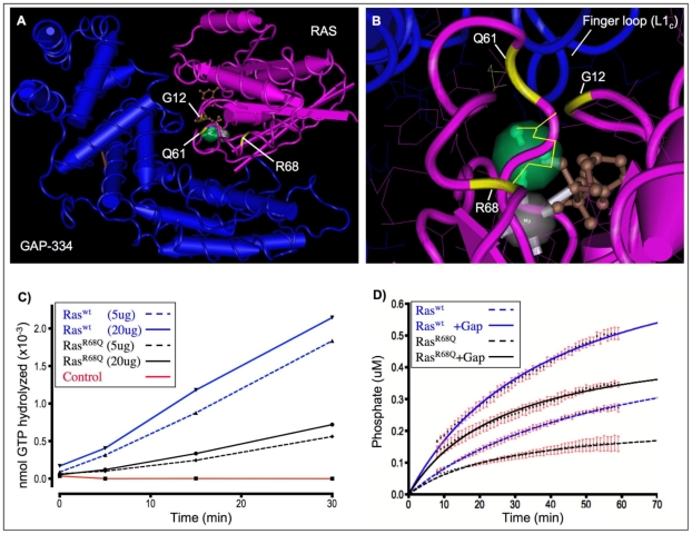
The RAS/MAPK signal transduction pathway is an intracellular signaling cascade that transmits environmental signals from activated receptor tyrosine kinases (RTKs) on the cell surface and other endomembranes to transcription factors in the nucleus, thereby linking extracellular stimuli to changes in gene expression. Largely as a consequence of its role in oncogenesis, RAS signaling has been the subject of intense research efforts for many years. More recently, it has been shown that milder perturbations in Ras signaling during embryogenesis also contribute to the etiology of a group of human diseases. Here we report the identification and characterization of the first gain-of-function germline mutation in Drosophila ras1 (ras85D), the Drosophila homolog of human K-ras, N-ras and H-ras. A single amino acid substitution (R68Q) in the highly conserved switch II region of Ras causes a defective protein with reduced intrinsic GTPase activity, but with normal sensitivity to GAP stimulation. The ras1(R68Q) mutant is homozygous viable but causes various developmental defects associated with elevated Ras signaling, including cell fate changes and ectopic survival of cells in the nervous system. These biochemical and functional properties are reminiscent of germline Ras mutants found in patients afflicted with Noonan, Costello or cardio-facio-cutaneous syndromes. Finally, we used ras1(R68Q) to identify novel genes that interact with Ras and suppress cell death.


Conflict of interest statement

Competing Interests: The authors have declared that no competing interests exist.

Figures

Figure 1
Figure 1. Suppression phenotypes of Su(21-3s).
GMR-hid but not GMR-grim or GMR-rpr induced cell death is dominantly suppressed by Su(21-3s) in a manner that requires intact MAPK phosphorylation sites in the overexpressed Hid protein. (A–F) The resulting rough eye phenotype is strongly suppressed in a dosage dependent manner by one (') or two (″) copies of the Su(21-3s) mutation when induced by overexpression of either a weak, GMR-hid1M (A) or a strong, GMR-hid10 (B) allele of GMR-hid, but is only weakly attenuated by Su(21-3s) when induced by GMR-grim (E) or GMR-rpr (F). In addition, Su(21-3s) suppresses cell death induced by overexpression of a Hid protein lacking 3 of 5 predicted MAPK phosphorylation sites, GMR-hidAla3 (C) but not by GMR-hidAla5 (D), a hid allele lacking all 5 MAPK consensus sites (Bergmann, et al. 1998). (G-I) Death of larval hemocytes induced by overexpression of Hid under control of the hemocyte specific driver Hml is also partially suppressed by the Su(21-3s) mutation. (G) EGFP is used to visualize hemocytes in wildtype 3rd instar larva: Hml-GAL4, 2xUAS-EGFP. (H) Overexpression of Hid in hemocytes results in their complete ablation by the 1st instar larval stage: Hml-Gal4, 2xUAS-EGFP; UAS-hid. (I) Su(21-3s) is able to partially suppress hemocyte death induced by Hid. Surviving hemocytes appear to be concentrated within the lymph glands as shown in the inset: Hml-Gal4, 2xUAS-EGFP; UAS-hid, Su(21-3s).
Figure 2
Figure 2. Su(21-3s) is a gain of function allele of ras1 (ras85D), the Drosophila homolog of human n-ras, h-ras and k-ras.
(A) The cell death suppression phenotype of Su(21-3s) mutants was localized by meiotic recombination to the right arm of the 3rd chromosome as indicated by the large horizontal arrow. P-element induced male recombination mapping further localized this suppressor to the region depicted by the short arrow. An enlargement of this interval (5.162-5.452 Mb on the physical map) is shown below, illustrating the ORFs contained therein. The ras85D (ras1) locus, outlined with a red box, was sequenced in a candidate gene approach and a G to A transition in exon3 (G641A) was identified. (B,C) A screen for reversion of the dominant Su(21-3s) suppressor phenotype generated a number of genetic revertants, two of which, Su(21-3s)R11 and Su(21-3s)41, were molecularly determined to be intragenic loss of function ras1 alleles. (B) A schematic of the ras1 locus, with exons boxed and coding regions stippled, illustrates the Su(21-3s) point mutation in exon 3 (G641A) and the small intragenic deletions identified in Su(21-3s)R11 and Su(21-3s)R41 (labeled R11 and R41 respectively, with deleted sequences underlined in black.) The red arrows correspond to primers used in a PCR diagnostic (below) used to confirm that both revertants contain the original G641A mutation. (C) Sequence analysis of these lethal revertants using strand specific PCR revealed that Su(21-3s)R11 contains an 18bp deletion that removes essential amino acids 87–92 from the Ras1 protein. Su(21-3s)R41 was found to have a 31bp deletion, resulting in a frameshift that generates a truncated Ras1 protein.
Figure 3
Figure 3. Amino acid alignment of fly, worm and mammalian Ras1.
These homologs have extensive primary sequence homology. Drosophila Ras1 (dmRas1), for example, is 87% identical to human K-ras (hsKras) at the amino acid level when C-terminal membrane-targeting sequences are excluded. Conserved regions are shaded in grey, with residues identical to the consensus sequence represented by a grey dot, while non-conserved residues remain unshaded. Five highly conserved signature motifs named “G box” sequences, labeled G1-G5 and boxed in red, are found in all families of small GTPases. Secondary structural elements are depicted as rectangles below the primary sequence alignment (alpha-helices, α1-α5, are dark grey and ß-sheets, ß1–ß6, light grey) and the phosphate-binding loop (P-loop), which binds the γamma-phosphate of GTP, and the nucleotide-sensitive switchI and II regions are indicated. The Switch regions are known to undergo large conformational changes upon exchange of bound GDP for GTP. The mutational spectrum of Ras is illustrated above the alignment, showing the distribution of amino acid substitutions encoded by germline mutations found for the developmental disorders Noonan, Costello and CFC syndromes and the most frequent cancer-associated somatic mutations (labeled in red). R68Q indicates the mutation characterized in this study, a non-conserved arginine to glutamine amino acid substitution within the switch II region of Drosophila Ras1 (dashed red box). hs, H. sapiens; dm, D. melanogaster; mm, M. musculus; ce, C. elegans.
Figure 4
Figure 4. Structural and biochemical analysis of wildtype and mutant Ras1.
(A–B) Three-dimensional crystal structure of human H-Ras (pink) bound to the GTPase-activating domain of human GTPase-activating protein p120GAP (GAP-334, blue) in the presence of aluminum fluoride (AlF3, green.) The positions of oncogenic residues glycine-12 (G12) and glutamine-61 (Q61) as well as the mutant residue in ras1R68Q flies, arginine-68 (R68), are shown in yellow. The Switch II region of Ras, of which Q61 and R68 are a part, is stabilized by GAP-334. (B) An enlargement of (A) showing the finger loop of GAP-334, which supplies an arginine side chain (arginine-789) into the active site of Ras to neutralize developing charges in the transition state (Scheffzek et al., 1997). R68, located proximally to the catalytic site of Ras, also extends a positively charged guanidinium group towards the active site. The images were constructed using the Entrez software Cn3D with mmdbId:51925 (Chen et al., 2003). Guanosine diphosphate (GDP,brown); Mg2+ (grey). (C) The intrinsic GTPase activities of affinity purified drosophila Ras1wt (blue) and Ras1R68Q (black) were determined using a kinetic phosphate assay employing [γamma-33P]GTP as a substrate. The conditions of the assay are such that the reaction proceeds with unimolecular kinetics and is insensitive to the amount of Ras protein employed (dashed vs. undashed lines). The mutant Ras1R68Q has an intrinsic GTPase activity that is approximately 1/3 that of wildtype Ras1 (kcat = 0.020 min−1 and 0.063 min−1 respectively.) (D) Human GAP-285 protein was purified by affinity chromatography and its ability to stimulate wildtype and mutant Drosophila Ras1 proteins was tested using a real-time fluorescent assay. Both wildtype and mutant Ras1 proteins are sensitive to GAP stimulation (dashed vs. undashed lines). Data is the average of three independent experiments. Error bars are in red.
Figure 5
Figure 5. Ras1R68Q mutants exhibit several developmental defects characteristic of elevated Ras/MAPK signaling.
(A–D) Semi-thin plastic sections of adult eyes were prepared and analyzed for defects in ommatidia formation. (A) Wildtype ommatidia are precisely ordered, containing one R7 cell and six outer photoreceptor cells. (B–D) Ras1R68Q ommatidia show two defects typical of mutations that increase Ras/MAPK signaling during eye development; supernumery R7 cells (arrows, B,C) and mislocalized (red circle, C) or missing (red circle, D) outer photoreceptor cells. The schematic illustrates the major cell types present in ommatidia. (E-G) Midline glial (MG) cells were visualized in wildtype (E) and ras1R68Q (F) stage 17 embryos using the MG-specific reporter construct P[slit-1.0-lacZ]. During development, the majority of MG undergo apoptosis such that at this stage only about three MG per segment normally survive. Elevated Ras/MAPK signaling allows for increased survival of MG cells and is reflected by an increase in the number of persisting MG cells per segment (arrows and inset, F). (G) Wildtype embryos contain an average of 2.8 MG cells per segment (n = 448) whereas ras1R68Q embryos contain an average of 3.3 MG cells per segment (n = 420). This difference is statistically significant by an unpaired t-test (p95≤0.0001). (H,I) Flies bearing the ras1R68Q allele develop ectopic wing material including extra longitudinal ‘veinlets’ near the posterior crossvein and an extra crossvein near the wing hinge (arrows, I) The area boxed in red is shown magnified below. PCV, posterior crossvein; ACV, anterior crossvein; L5, L5 wing vein.

Similar articles

Cited by

References

    1. Cantley LC, Auger KR, Carpenter C, Duckworth B, Graziani A, et al. Oncogenes and signal transduction. Cell. 1991;64:281–302. - PubMed
    1. Blume-Jensen P, Hunter T. Oncogenic kinase signalling. Nature. 2001;411:355–365. - PubMed
    1. Bishop JM. Molecular themes in oncogenesis. Cell. 1991;64:235–248. - PubMed
    1. Hunter T. Cooperation between oncogenes. Cell. 1991;64:249–270. - PubMed
    1. Colicelli J. Vol. 2004. Sci STKE; 2004. Human RAS superfamily proteins and related GTPases.RE13 - PMC - PubMed

Publication types

MeSH terms