. 2011;6(8):e23540.
doi: 10.1371/journal.pone.0023540. Epub 2011 Aug 17.

Toll-like receptor 2 induced angiogenesis and invasion is mediated through the Tie2 signalling pathway in rheumatoid arthritis

Affiliations

Toll-like receptor 2 induced angiogenesis and invasion is mediated through the Tie2 signalling pathway in rheumatoid arthritis

Tajvur Saber et al. PLoS One. 2011.

Abstract

Background: Angiogenesis is a critical early event in inflammatory arthritis, facilitating leukocyte migration into the synovium resulting in invasion and destruction of articular cartilage and bone. This study investigates the effect of TLR2 on angiogenesis, EC adhesion and invasion using microvascular endothelial cells and RA whole tissue synovial explants ex-vivo.

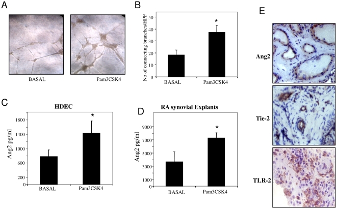
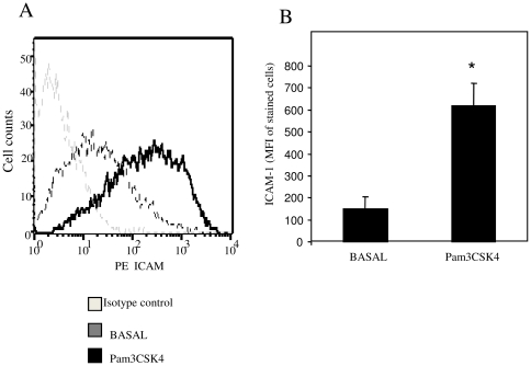
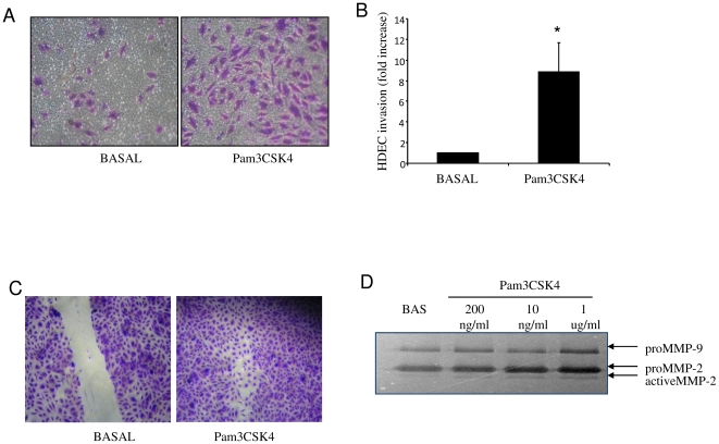
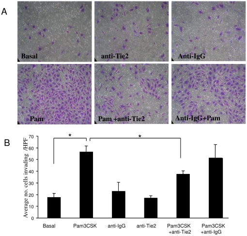
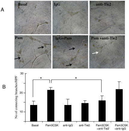
Methods: Microvascular endothelial cells (HMVEC) and RA synovial explants ex vivo were cultured with the TLR2 ligand, Pam3CSK4 (1 µg/ml). Angiopoietin 2 (Ang2), Tie2 and TLR2 expression in RA synovial tissue was assessed by immunohistology. HMVEC tube formation was assessed using Matrigel matrix assays. Ang2 was measured by ELISA. ICAM-1 cell surface expression was assessed by flow cytometry. Cell migration was assessed by wound repair scratch assays. ECM invasion, MMP-2 and -9 expression were assessed using transwell invasion chambers and zymography. To examine if the angiopoietin/Tie2 signalling pathway mediates TLR2 induced EC tube formation, invasion and migration assays were performed in the presence of a specific neutralising anti-Tie2mAb (10 ug/ml) and matched IgG isotype control Ab (10 ug/ml).

Results: Ang2 and Tie2 were localised to RA synovial blood vessels, and TLR2 was localised to RA synovial blood vessels, sub-lining infiltrates and the lining layer. Pam3CSK4 significantly increased angiogenic tube formation (p<0.05), and upregulated Ang2 production in HMVEC (p<0.05) and RA synovial explants (p<0.05). Pam3CSK4 induced cell surface expression of ICAM-1, from basal level of 149±54 (MFI) to 617±103 (p<0.01). TLR-2 activation induced an 8.8±2.8 fold increase in cell invasion compared to control (p<0.05). Pam3CSK4 also induced HMVEC cell migration and induced MMP-2 and -9 from RA synovial explants. Neutralisation of the Ang2 receptor, Tie2 significantly inhibited Pam3CSK4-induced EC tube formation and invasion (p<0.05).

Conclusion: TLR2 activation promotes angiogenesis, cell adhesion and invasion, effects that are in part mediated through the Tie2 signalling pathway, key mechanisms involved in the pathogenesis of RA.


Conflict of interest statement

Competing Interests: The authors have declared that no competing interests exist.

Figures

Figure 1
Figure 1. TLR2 activation induces EC tube formation and Ang2 expression.
Human dermal microvascular endothelial cell tubule formation on matrigel matrix following stimulation with Pam3CSK4 (1 ug/ml). (A) Representative image of baseline tube formation (left panel) and tube formation following stimulation with Pam3CSK4 (right panel). (B) Quantitative analysis of the number of connecting branches at baseline and in response to Pam3CSK4. The tube analysis was determined from 5 sequential fields (Magnification×40) focussing on the surface of the matrigel (n = 4). (C–D) The effect of Pam3CSK4 on angiopoietin-2 expression in HMVEC (n = 4) and RA synovial explants (n = 6). Data represented as the mean+/−sem. *p<0.05 significantly different from baseline. (E) Ang2, Tie2 and TLR2 expression in RA synovial tissue sections.
Figure 2
Figure 2. TLR2 activation induces ICAM-1 cell surface expression HMVEC.
Human dermal microvascular endothelial cells were stimulated with Pam3CSK4 (1 ug/ml) and expression of ICAM-1 was detected by flow cytometric analysis. (A) Representative flow cytometry histogram demonstrating induced ICAM-1 expression on HMVEC following stimulation with Pam3CSK4 (black line) compared to basal (grey line). B. Quantification of ICAM-1 expression following incubation with Pam3CSK4. Data represented as mean fluorescent intensity (mean±sem, n = 4). * p<0.05 significantly different from unstimulated.
Figure 3
Figure 3. TLR-2 activation induces EC invasion and migration and MMP-2 and 9 expression in RA synovial explants.
(A) Representative photomicrograph shows HMVEC invasion following Pam3CSK4 (1 µg/ml), stimulation. At 24 hours invading cells attached to lower membrane were fixed (1% glutaraldehyde) and stained (0.1% crystal violet) (Mag×40). (B) Representative bar graph quantifying HMVEC invasion. (n = 4). *p<0.05 significantly different to control. (C) Representative photomicrograph showing cells repopulating the wound in response to Pam3CSK4 (1 ug/ml). (D) Representative gel of MMP-2 and 9 activity by gelatine zymography in response to Pam3CSK4 in RA synovial explants (n = 3).
Figure 4
Figure 4. TLR2 induced EC invasion is inhibited by anti-Tie2.
(A) Representative photomicrographs showing anti-Tie2 blocks Pam3CSK4 induced HMVEC invasion, with no effect observed for IgG control mAb. At 24 hours invading cells attached to lower membrane were fixed (1% glutaraldehyde) and stained (0.1% crystal violet) (Mag×40). (B) Representative bar graph quantifying HMVEC invasion. (n = 4). *p<0.05 significantly different.
Figure 5
Figure 5. TLR2 induced EC tube formation and wound repair are blocked by anti-Tie2.
(A) Representative photomicrographs showing anti-Tie2 blockade of TLR-2 induced angiogenesis, with effect observed for IgG control mAb (n = 3 experiments). Black arrows indicates an increase in EC-EC connecting branches and tube formation in response to PAM3CSK4, white arrow indicates a decrease in the number of connecting branches in the presence of an-Tie2. (B) Representative bar graph quantifying HMVEC tube formation *p<0.05 significantly different.

References

    1. Koch AE. Angiogenesis as a target in rheumatoid arthritis. Ann Rheum Dis. 2003;62:60–67. - PMC - PubMed
    1. Ferrara N, Davis-Smyth T. The biology of vascular endothelial growth factor. Endocr Rev. 1997;18:4–25. - PubMed
    1. Bucala R, Ritchlin C, Winchester R, Cerami A. Constitutive production of inflammatory and mitogenic cytokines by rheumatoid synovial fibroblasts. J Exp Med. 1991;173:569–574. - PMC - PubMed
    1. Chu CQ, Field M, Allard S, Abney E, Feldmann M, et al. Detection of cytokines at the cartilage/pannus junction in patients with rheumatoid arthritis: implications for the role of cytokines in cartilage destruction and repair. Br J Rheumatol. 1992;10:653–661. - PubMed
    1. Tak PP, Bresnihan B. The pathogenesis and prevention of joint damage in rheumatoid arthritis: advances from synovial biopsy and tissue analysis. Arthritis Rheum. 2000;43:2619–2633. - PubMed

Publication types

MeSH terms