

The distribution of phosphatidylinositol 4,5-bisphosphate in acinar cells of rat pancreas revealed with the freeze-fracture replica labeling method
- PMID: 21858170
- PMCID: PMC3156236
- DOI: 10.1371/journal.pone.0023567
The distribution of phosphatidylinositol 4,5-bisphosphate in acinar cells of rat pancreas revealed with the freeze-fracture replica labeling method
Abstract
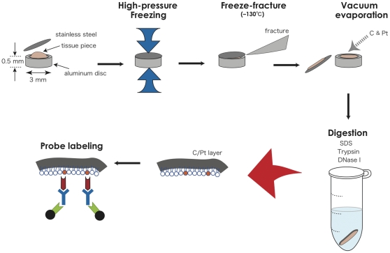
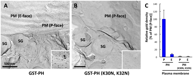
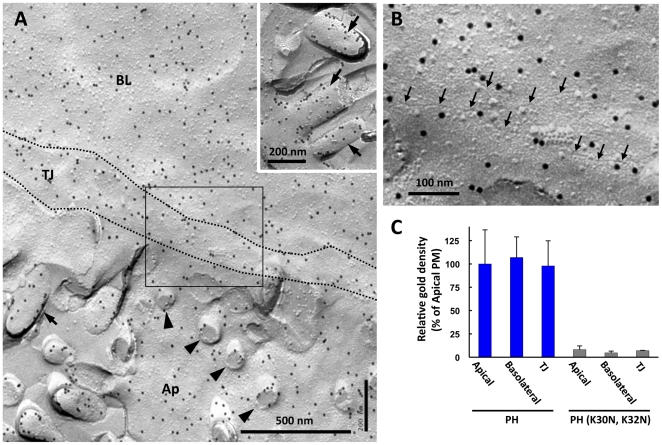
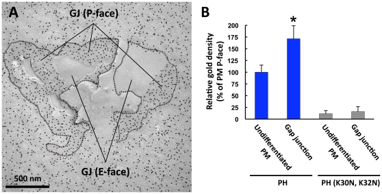
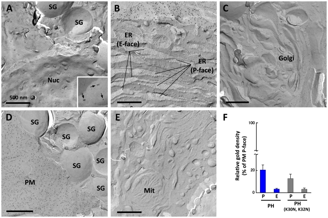
Phosphatidylinositol 4,5-bisphosphate [PI(4,5)P(2)] is a phospholipid that has been implicated in multiple cellular activities. The distribution of PI(4,5)P(2) has been analyzed extensively using live imaging of the GFP-coupled phospholipase C-δ1 pleckstrin homology domain in cultured cell lines. However, technical difficulties have prevented the study of PI(4,5)P(2) in cells of in vivo tissues. We recently developed a method to analyze the nanoscale distribution of PI(4,5)P(2) in cultured cells by using the quick-freezing and freeze-fracture replica labeling method. In principle, this method can be applied to any cell because it does not require the expression of artificial probes. In the present study, we modified the method to study cells of in vivo tissues and applied it to pancreatic exocrine acinar cells of the rat. We found that PI(4,5)P(2) in the plasma membrane is distributed in an equivalent density in the apical and basolateral domains, but exists in a significantly higher concentration in the gap junction. The intracellular organelles did not show labeling for PI(4,5)P(2). The results are novel or different from the reported distribution patterns in cell lines and highlight the importance of studying cells differentiated in vivo.
Conflict of interest statement
Figures
Similar articles
-
A method for efficient observation of intracellular membranes of monolayer culture cells by quick-freeze and freeze-fracture electron microscopy.J Electron Microsc (Tokyo). 2012;61(6):441-6. doi: 10.1093/jmicro/dfs063. Epub 2012 Nov 25. J Electron Microsc (Tokyo). 2012. PMID: 23183965
-
Fine structural analysis of a teleost exocrine pancreas cellular components - a freeze-fracture and transmission electron microscopic study.Anat Anz. 1980;147(1):60-75. Anat Anz. 1980. PMID: 7396226
-
Ultrastructural aspects of acute pancreatitis induced by 2, 2'-azobis (2-amidinopropane) dihydrochloride (AAPH) in rats.Folia Morphol (Warsz). 2012 Aug;71(3):136-41. Folia Morphol (Warsz). 2012. PMID: 22936547
-
Gap junctions revealed by freeze-fracture electron microscopy.Microsc Res Tech. 1995 Aug 1;31(5):437-45. doi: 10.1002/jemt.1070310512. Microsc Res Tech. 1995. PMID: 8534904 Review.
-
SDS-digested freeze-fracture replica labeling electron microscopy to study the two-dimensional distribution of integral membrane proteins and phospholipids in biomembranes: practical procedure, interpretation and application.Histochem Cell Biol. 1997 Feb;107(2):87-96. doi: 10.1007/s004180050092. Histochem Cell Biol. 1997. PMID: 9062793 Review.
Cited by
-
Long-term depression in neurons involves temporal and ultra-structural dynamics of phosphatidylinositol-4,5-bisphosphate relying on PIP5K, PTEN and PLC.Commun Biol. 2023 Apr 3;6(1):366. doi: 10.1038/s42003-023-04726-0. Commun Biol. 2023. PMID: 37012315 Free PMC article.
-
Polarity and stratification of the epidermis.Semin Cell Dev Biol. 2012 Oct;23(8):890-6. doi: 10.1016/j.semcdb.2012.08.008. Epub 2012 Aug 31. Semin Cell Dev Biol. 2012. PMID: 22960184 Free PMC article. Review.
-
Polarized PtdIns(4,5)P2 distribution mediated by a voltage-sensing phosphatase (VSP) regulates sperm motility.Proc Natl Acad Sci U S A. 2019 Dec 17;116(51):26020-26028. doi: 10.1073/pnas.1916867116. Epub 2019 Nov 27. Proc Natl Acad Sci U S A. 2019. PMID: 31776261 Free PMC article.
-
PDK1 in apical signaling endosomes participates in the rescue of the polarity complex atypical PKC by intermediate filaments in intestinal epithelia.Mol Biol Cell. 2012 May;23(9):1664-74. doi: 10.1091/mbc.E11-12-0988. Epub 2012 Mar 7. Mol Biol Cell. 2012. PMID: 22398726 Free PMC article.
-
Phosphoinositides: tiny lipids with giant impact on cell regulation.Physiol Rev. 2013 Jul;93(3):1019-137. doi: 10.1152/physrev.00028.2012. Physiol Rev. 2013. PMID: 23899561 Free PMC article. Review.
References
-
- Di Paolo G, De Camilli P. Phosphoinositides in cell regulation and membrane dynamics. Nature. 2006;443:651–657. - PubMed
-
- Downes CP, Gray A, Lucocq JM. Probing phosphoinositide functions in signaling and membrane trafficking. Trends Cell Biol. 2005;15:259–268. - PubMed
-
- Irvine R. Inositol lipids: to PHix or not to PHix? Curr Biol. 2004;14:R308–310. - PubMed
-
- Gillooly DJ, Raiborg C, Stenmark H. Phosphatidylinositol 3-phosphate is found in microdomains of early endosomes. Histochem Cell Biol. 2003;120:445–453. - PubMed
Publication types
MeSH terms
Substances
LinkOut - more resources
Full Text Sources
Research Materials
Miscellaneous
