

Dynamic changes in EPCAM expression during spermatogonial stem cell differentiation in the mouse testis
- PMID: 21858196
- PMCID: PMC3156235
- DOI: 10.1371/journal.pone.0023663
Dynamic changes in EPCAM expression during spermatogonial stem cell differentiation in the mouse testis
Abstract
Background: Spermatogonial stem cells (SSCs) have the unique ability to undergo self-renewal division. However, these cells are morphologically indistinguishable from committed spermatogonia, which have limited mitotic activity. To establish a system for SSC purification, we analyzed the expression of SSC markers CD9 and epithelial cell adhesion molecule (EPCAM), both of which are also expressed on embryonic stem (ES) cells. We examined the correlation between their expression patterns and SSC activities.
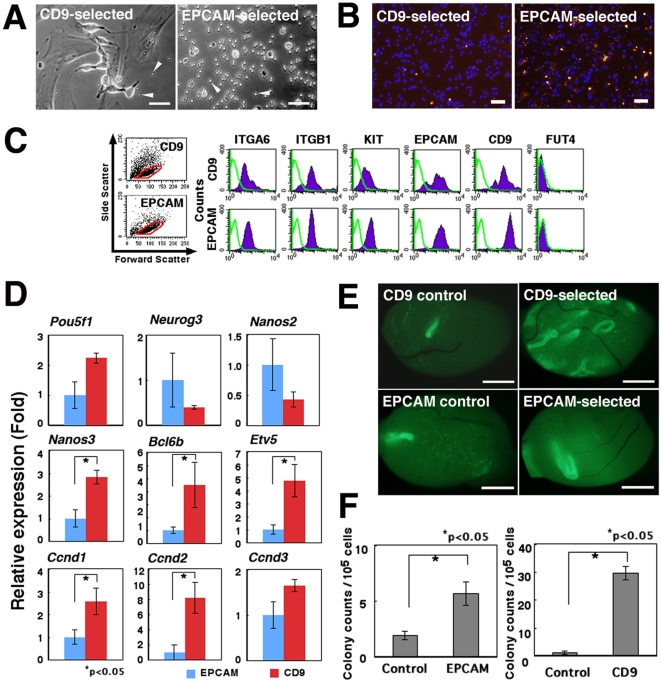
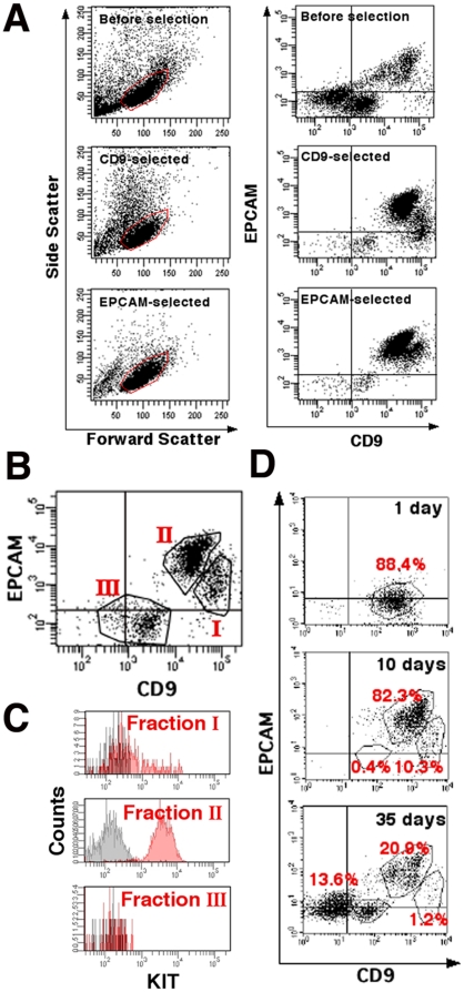
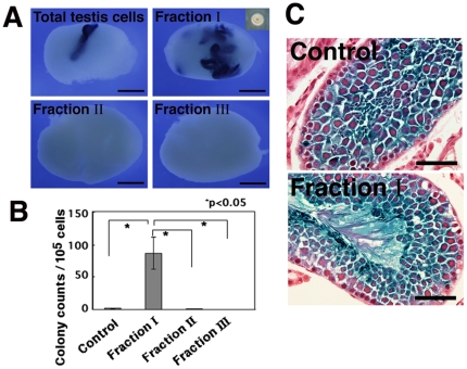
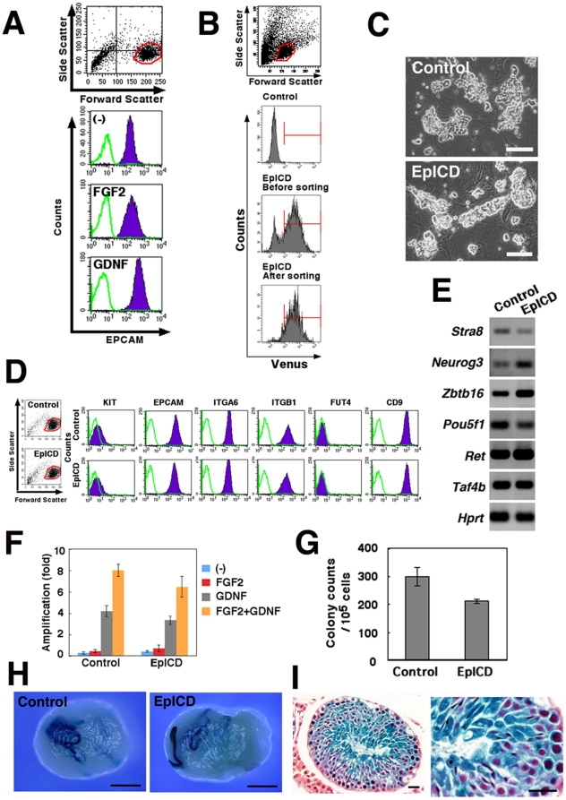
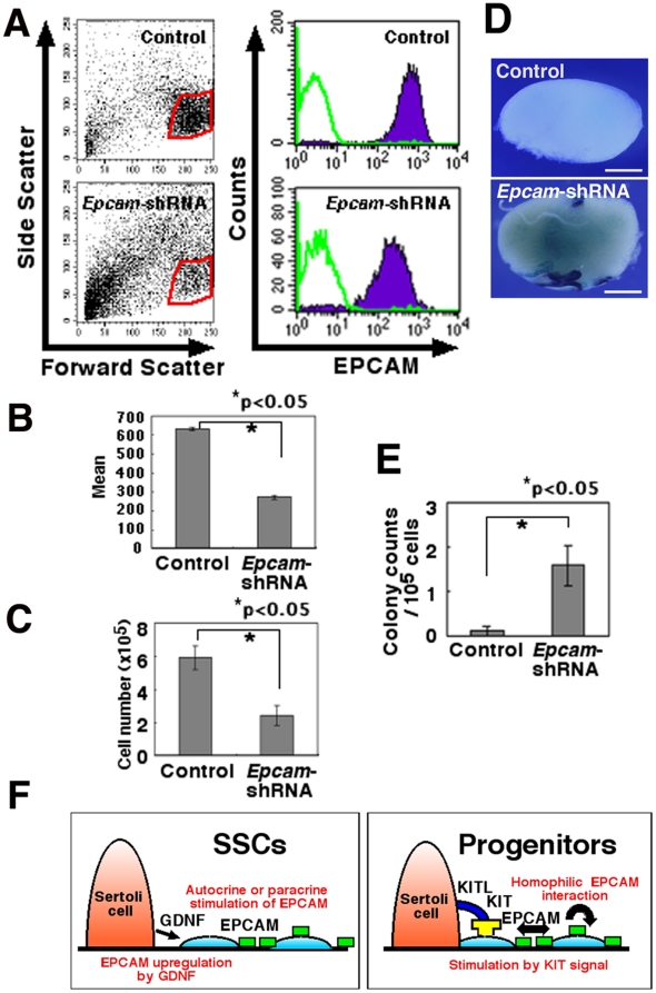
Methodology and principal findings: By magnetic cell sorting, we found that EPCAM-selected mouse germ cells have limited clonogenic potential in vitro. Moreover, these cells showed stronger expression of progenitor markers than CD9-selected cells, which are significantly more enriched in SSCs. Fluorescence-activated cell sorting of CD9-selected cells indicated a significantly higher frequency of SSCs among the CD9(+)EPCAM(low/-) population than among the CD9(+)EPCAM(+) population. Overexpression of the active form of EPCAM in germline stem (GS) cell cultures did not significantly influence SSC activity, whereas EPCAM suppression by short hairpin RNA compromised GS cell proliferation and increased the concentration of SSCs, as revealed by germ cell transplantation.
Conclusions/significance: These results show that SSCs are the most concentrated in CD9(+)EPCAM(low/-) population and also suggest that EPCAM plays an important role in progenitor cell amplification in the mouse spermatogenic system. The establishment of a method to distinguish progenitor spermatogonia from SSCs will be useful for developing an improved purification strategy for SSCs from testis cells.
Conflict of interest statement
Figures
Similar articles
-
Enrichment of mouse spermatogonial stem cells by melanoma cell adhesion molecule expression.Biol Reprod. 2012 Dec 13;87(6):139. doi: 10.1095/biolreprod.112.103861. Print 2012 Jun. Biol Reprod. 2012. PMID: 23053437
-
Fluorescence- and magnetic-activated cell sorting strategies to isolate and enrich human spermatogonial stem cells.Fertil Steril. 2014 Aug;102(2):566-580.e7. doi: 10.1016/j.fertnstert.2014.04.036. Epub 2014 Jun 2. Fertil Steril. 2014. PMID: 24890267 Free PMC article.
-
MicroRNA-21 regulates the self-renewal of mouse spermatogonial stem cells.Proc Natl Acad Sci U S A. 2011 Aug 2;108(31):12740-5. doi: 10.1073/pnas.1109987108. Epub 2011 Jul 18. Proc Natl Acad Sci U S A. 2011. PMID: 21768389 Free PMC article.
-
Mechanisms regulating mammalian spermatogenesis and fertility recovery following germ cell depletion.Cell Mol Life Sci. 2019 Oct;76(20):4071-4102. doi: 10.1007/s00018-019-03201-6. Epub 2019 Jun 28. Cell Mol Life Sci. 2019. PMID: 31254043 Free PMC article. Review.
-
Spermatogonial stem cell transplantation and testicular function.Cell Tissue Res. 2005 Oct;322(1):21-31. doi: 10.1007/s00441-005-0009-z. Epub 2005 Nov 3. Cell Tissue Res. 2005. PMID: 16047158 Review.
Cited by
-
Effects of transgene insertion loci and copy number on Dnmt3L gene silencing through antisense transgene-derived PIWI-interacting RNAs.RNA. 2022 May;28(5):683-696. doi: 10.1261/rna.078905.121. Epub 2022 Feb 10. RNA. 2022. PMID: 35145000 Free PMC article.
-
Purification of GFRα1+ and GFRα1- Spermatogonial Stem Cells Reveals a Niche-Dependent Mechanism for Fate Determination.Stem Cell Reports. 2018 Feb 13;10(2):553-567. doi: 10.1016/j.stemcr.2017.12.009. Epub 2018 Jan 11. Stem Cell Reports. 2018. PMID: 29337115 Free PMC article.
-
Expression of Genes Related to Germ Cell Lineage and Pluripotency in Single Cells and Colonies of Human Adult Germ Stem Cells.Stem Cells Int. 2016;2016:8582526. doi: 10.1155/2016/8582526. Epub 2015 Nov 8. Stem Cells Int. 2016. PMID: 26649052 Free PMC article.
-
Distinctive molecular features of regenerative stem cells in the damaged male germline.Nat Commun. 2022 May 6;13(1):2500. doi: 10.1038/s41467-022-30130-z. Nat Commun. 2022. PMID: 35523793 Free PMC article.
-
Effect of Seminal Plasma on the Freezability of Boar Sperm.Animals (Basel). 2024 Dec 18;14(24):3656. doi: 10.3390/ani14243656. Animals (Basel). 2024. PMID: 39765560 Free PMC article.
References
-
- de Rooij DG, Russell LD. All you wanted to know about spermatogonia but were afraid to ask. J Androl. 2000;21:776–798. - PubMed
-
- Meistrich ML, van Beek MEAB. Spermatogonial stem cells. In: Desjardins CC, Ewing LL, editors. Cell and Molecular Biology of the Testis. New York: Oxford University Press; 1993. pp. 266–295.
-
- Tegelenbosch RAJ, de Rooij DG. A quantitative study of spermatogonial multiplication and stem cell renewal in the C3H/101 F1 hybrid mouse. Mutation Res. 1993;290:193–200. - PubMed
Publication types
MeSH terms
Substances
LinkOut - more resources
Full Text Sources
Medical
Miscellaneous
