. 2011;6(8):e23817.
doi: 10.1371/journal.pone.0023817. Epub 2011 Aug 17.

Regulation of anthrax toxin-specific antibody titers by natural killer T cell-derived IL-4 and IFNγ

Affiliations

Regulation of anthrax toxin-specific antibody titers by natural killer T cell-derived IL-4 and IFNγ

T Scott Devera et al. PLoS One. 2011.

Abstract

Activation of Natural Killer-like T cells (NKT) with the CD1d ligand α-GC leads to enhanced production of anthrax toxin protective Ag (PA)-neutralizing Abs, yet the underlying mechanism for this adjuvant effect is not known. In the current study we examined the role of Th1 and Th2 type responses in NKT-mediated enhancement of antibody responses to PA. First, the contribution of IL-4 and IFNγ to the production of PA-specific toxin-neutralizing Abs was examined. By immunizing C57Bl/6 controls IL-4(-/-) mice and IFNγ(-/-) mice and performing passive serum transfer experiments, it was observed that sera containing PA-specific IgG1, IgG2b and IgG2c neutralized toxin in vitro and conferred protection in vivo. Sera containing IgG2b and IgG2c neutralized toxin in vitro but were not sufficient for protection in vivo. Sera containing IgG1 and IgG2b neutralized toxin in vitro and conferred protection in vivo. IgG1 therefore emerged as a good correlate of protection. Next, C57Bl/6 mice were immunized with PA alone or PA plus a Th2-skewing α-GC derivative known as OCH. Neutralizing PA-specific IgG1 responses were modestly enhanced by OCH in C57Bl/6 mice. Conversely, IgG2b and IgG2c were considerably enhanced in PA/OCH-immunized IL-4(-/-) mice but did not confer protection. Finally, bone marrow chimeras were generated such that NKT cells were unable to express IL-4 or IFNγ. NKT-derived IL-4 was required for OCH-enhanced primary IgG1 responses but not recall responses. NKT-derived IL-4 and IFNγ also influenced primary and recall IgG2b and IgG2c titers. These data suggest targeted skewing of the Th2 response by α-GC derivatives can be exploited to optimize anthrax vaccination.


Conflict of interest statement

Competing Interests: The authors have declared that no competing interests exist.

Figures

Figure 1
Figure 1. Requirement for IL-4 and IFNγ in production of PA-specific IgG1 or IgG2c.
C57Bl/6, IL-4−/− and IFNγ−/− mice were immunized and bled as described. A Endpoint anti-PA IgG1, B IgG2b, and C IgG2c titers were determined by ELISA. Each data point represents an individual mouse and geometric mean titers are shown. Data are representative of three independent experiments. Statistical significance (p<0.05) is indicated by asterisks.
Figure 2
Figure 2. Requirement for IL-4 but not IFNγ in production of PA-neutralizing IgG.
A Sera from experiment in Figure 1 were tested for their ability to protect RAW267.4 macrophages from LT toxicity. Graph shows neutralization by sera from C57Bl/6 mice, IL-4−/− and IFNγ−/− mice. Each data point represents an individual mouse and the mean survival of the cell line following treatment with LT and sera is indicated. B Mice were challenged with LT as indicated. C Pooled sera from naïve (n = 5) and PA-immunized (n = 5) mice were transferred to naïve IL-4−/− mice before challenge with LT. Three and four IL-4−/− mice per group received naïve and immune sera respectively. Data in A and B are representative of two independent experiments while data in C are from a single experiment. Graphs show percent survival following LT administration. Statistical significance (p<0.05) is indicated by asterisks.
Figure 3
Figure 3. Effect of OCH on PA-specific IgG1, IgG2b and IgG2c response.
C57Bl/6 mice were immunized and bled as described. A Endpoint anti-PA IgG1, B IgG2b and C IgG2c titers were determined by ELISA. Each data point represents an individual mouse (5 per group) and geometric mean titers are shown. Data are representative of two independent experiments. Statistical significance (p<0.05) is indicated by asterisks.
Figure 4
Figure 4. OCH-enhanced IgG2b and IgG2c responses in IL-4−/− mice.
C57Bl/6 and IL-4−/− mice were immunized and bled as described in materials and methods. A Shows endpoint anti-PA IgG1 titers. B Shows endpoint anti-PA IgG2b titers. C Shows endpoint anti-IgG2c titers. Each data point represents an individual mouse (4 or 5 per group as indicated) and geometric mean titers are shown. Data are representative of two independent experiments.
Figure 5
Figure 5. Non-protective OCH-enhanced IgG2b and IgG2c responses in IL-4−/− mice.
A Sera from mice in Figure 4 were tested for neutralization of LT in vitro. B C57Bl/6 and C IL-4−/− mice were challenged as indicated. Toxin was administered on d 50 when IgG2 titers were highest. Graphs show percent survival following LT administration. Statistical significance (p<0.05) is indicated by asterisks. Each data point in A represents an individual mouse (4 or 5 per group as indicated). All mice in A were challenged for parts B and C. Data are representative of two independent experiments.
Figure 6
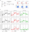
Figure 6. Modulation of PA-specific IgG1, IgG2b and IgG2c by NKT-derived IL-4 and IFNγ.
A Bone marrow chimeras were generated as indicated and detailed in materials and methods. B Splenocytes from chimeras were analyzed by flow cytometry for expression of CD1d (histograms) and re-constitution of CD1d-tetramer+/TCRβ+ NKT cells (dot plots). Mean fluorescent intensity for anti-CD1d mAb binding and percent frequency of CD1d-tetramer+/TCRβ+ cells are indicated. Comparable numbers of total splenocytes were recovered from each chimera indicating comparable absolute numbers of NKT cells. Reconstitution of cell populations and NKT cells has been demonstrated in at least 6 independent experiments. C Chimeric mice were immunized and bled as described. Top row shows endpoint anti-PA IgG1 titers on the days indicated following immunization. Middle row shows endpoint anti-PA IgG2b titers. Bottom row shows endpoint anti-IgG2c titers. Each data point represents an individual mouse (5–7 per group as indicated). Data are representative of two similar independent experiments (except for α-GC as adjuvant). Statistical significance (p<0.05) is indicated by asterisks. Further statistical comparisons (ANOVA) were employed to compare the d45 bleeds for IgG2b and IgG2c between the three different chimeras. The IgG2b and IgG2c titers were significantly lower in the Jα18−/−/IFNγ−/− chimeras than the Jα18−/−/IL-4−/− chimeras but not the Jα18−/−/C57Bl/6 chimeras (not depicted on figure).

References

    1. Bradley KA, Mogridge J, Mourez M, Collier RJ, Young JA. Identification of the cellular receptor for anthrax toxin. Nature. 2001;414:225–229. - PubMed
    1. Collier RJ, Young JA. Anthrax toxin. Annu Rev Cell Dev Biol. 2003;19:45–70. - PubMed
    1. Leppla SH. Bacillus anthracis calmodulin-dependent adenylate cyclase: chemical and enzymatic properties and interactions with eucaryotic cells. Adv Cyclic Nucleotide Protein Phosphorylation Res. 1984;17:189–198. - PubMed
    1. Fink SL, Bergsbaken T, Cookson BT. Anthrax lethal toxin and Salmonella elicit the common cell death pathway of caspase-1-dependent pyroptosis via distinct mechanisms. Proc Natl Acad Sci U S A. 2008;105:4312–4317. - PMC - PubMed
    1. Mock M, Mignot T. Anthrax toxins and the host: a story of intimacy. Cell Microbiol. 2003;5:15–23. - PubMed

Publication types

MeSH terms