. 2011;6(8):e23838.
doi: 10.1371/journal.pone.0023838. Epub 2011 Aug 17.

The hepatitis C virus E1 glycoprotein undergoes productive folding but accelerated degradation when expressed as an individual subunit in CHO cells

Affiliations

The hepatitis C virus E1 glycoprotein undergoes productive folding but accelerated degradation when expressed as an individual subunit in CHO cells

Valentina Botti et al. PLoS One. 2011.

Abstract

Hepatitis C Virus E1E2 heterodimers are components of the viral spike. Although there is a general agreement on the necessity of the co-expression of both E1 and E2 on a single coding unit for their productive folding and assembly, in a previous study using an in vitro system we obtained strong indications that E1 can achieve folding in absence of E2. Here, we have studied the folding pathway of unescorted E1 from stably expressing CHO cells, compared to the folding observed in presence of the E2 protein. A DTT-resistant conformation is achieved by E1 in both situations, consistent with the presence of an E2-independent oxidative pathway. However, while the E1E2 heterodimer is stable inside cells, E1 expressed alone is degraded within a few hours. On the other hand, the oxidation and stability of individually expressed E2 subunits is dependent on E1 co-expression. These data are consistent with E1 and E2 assisting each other for correct folding via different mechanisms: E2 assists E1 by stabilizing a semi-native conformation meanwhile E1 drives E2 towards a productive folding pathway.


Conflict of interest statement

Competing Interests: The authors have declared that no competing interests exist.

Figures

Figure 1
Figure 1. Endoglycosidase H sensitivity of HCV glycoproteins E1 and E2p7.
(A) Recombinant constructs for E1, E2p7 and E1E2p7 expression in CHO cells. Sequences coding for the structural protein E1 (aa. 165–398), E2p7 (aa. 365–809) and E1E2p7 (aa. 165–809) were amplified from a replicon containing the entire HCV genome of HCV genotype 1b, subtype ConI as described in the Materials and Methods. These constructs were used to stably express the proteins in CHO cells. Black segments represent the E1 (aa. 165–191) and E2 (aa. 366–384) signal sequences. The N-terminus 15 amino acids of the E2 protein are shown as a dashed segment in the E1 construct. (B) CHO cells expressing E1, E2p7 or E1E2p7 were pulse-labeled with [35S]-methionine and -cysteine for 20 min and chased for the indicated time periods. PNSs were immunoprecipitated with an anti-E1 (top panel) or anti-E2 antibody (lower panel) and further treated with (+) or without (−) EndoH. Proteins were separated on 10% SDS-PAGE in reducing conditions. Asterisks indicate non-specific bands. E1ΔGly indicates the E1 protein co-immunoprecipitated with the anti-E2 antibody and subsequentlydeglycosylated by EndoH treatment.
Figure 2
Figure 2. Oxidation kinetics of E1 and E2p7 HCV glycoprotein.
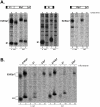
CHO cells stably transfected with constructs expressing E1, E2p7 or E1E2p7 were pulse-labeled with [35S]-methionine and -cysteine for 20 min and chased for the indicated time-periods. PNSs were immunoprecipitated with (A) CBH-111 anti-E1 monoclonal antibody or (B) CBH-7 anti-E2 monoclonal antibody. Samples were analyzed on 10% SDS-PAGE in non-reducing (lanes 1–5) and reducing (lanes 6–10) conditions. For E1, the relative intensity of the unoxidized (grey bars) and oxidized (black bars) forms were quantified for the different chase-times. The E1E2p7 wild type species is reported on the left graph, E1 alone on the right graph. The half-time (t1/2) of E1 oxidation represents the chase-time at which half of the protein is in its oxidized form. Symbols refer to: ox for oxidized; unox for unoxidized; red for reduced.
Figure 3
Figure 3. Analysis of DTT-resistance of HCV proteins E1 and E2.
(A) CHO cells stably transfected with E1E2p7 or E1 were pulse-labeled with [35S]-methionine and –cysteine for 20 min and chased for different time-periods in the presence (+) or absence (−) of 5 mM DTT added exogenously 5 min before the indicated chase-time. PNSs were immunoprecipitated with the CBH-111 anti-E1 antibody and samples analyzed on 10% SDS-PAGE in non-reducing (lanes 1–10) and reducing (lanes 11–20) conditions. Graphs below represent the percentage of oxidized E1 present in samples derived from untreated (black bars) and DTT-treated (dashed bars) cells. The relative intensity-percentage is calculated by densitometric analysis of the unoxidized and oxidized forms in non-reducing conditions in the presence (lanes 1, 3, 5, 7, 9) or absence (lanes 2, 4, 6, 8) of 5 mM DTT added exogenously 5 min before the indicated chase-period. Left graph reports values obtained when expressed with E2p7, right graph when expressed individually. (B) CHO cells stably transfected with E1E2p7 or E2p7 were pulse-labeled with [35S]-methionine and –cysteine for 20 min and chased for different time-periods in the presence (+) or absence (−) of 5 mM DTT added exogenously 5 min before the indicated chase-time. PNSs were immunoprecipitated with CBH-7 anti-E2 antibody and samples analyzed on 10% SDS-PAGE in non-reducing (lanes 1–5) and reducing (lanes 6–10) conditions. Symbols refer to: ox for oxidized; unox for unoxidized; red for reduced.
Figure 4
Figure 4. E1 and E2 stability in CHO stably trasfected cells.
(A) CHO cells expressing E1, E2p7 or E1E2p7 were pulse-labeled with [35S]-methionine and –cysteine for 20 min and chased for 6 and 15 hrs. PNSs were immunoprecipitated with the CBH-111 anti-E1 monoclonal antibody (central panel) or the CBH-7 anti-E2 monoclonal antibody (lateral panels). Samples were analyzed on 10% SDS-PAGE in non-reducing (lanes 1, 2, 5, 6, 9, 10) or reducing (lanes 3, 4, 7, 8, 11, 12) conditions. (B) CHO cells stably transfected with E1, E2p7 or E1E2p7 were pulse-labeled with [35S]-methionine and –cysteine for 20 min and 15 hrs. During the pulse- and chase-time, the medium was supplemented with (+) or without (−) 20 µM kifunesine. PNSs were immunoprecipitated with the CBH-111 anti-E1 antibody (lanes 3, 4 and 9, 10) or the CBH-7 anti-E2 monoclonal antibody (lanes 1, 2, 5, 6, 7, 8, 11, 12). Samples were analyzed on 10% SDS-PAGE in non-reducing (lanes 1–6) or reducing (lanes 7–12) conditions.

Similar articles

Cited by

References

    1. Lemon SM, Walker C, Alter MJ, Yi M. Hepatitis C Virus. In: Knipe DM, Howley PM, editors. Fields Virology. Philadelphia, PA, USA: Lippincott Williams & Wilkins; 2007. pp. 1253–1304.
    1. Lindenbach BD, Thiel HJ, Rice CM. Flaviviridae: the viruses and their replication. In: Knipe DM, Howley PM, editors. Fields Virology. Philadelphia, PA, USA: Lippincott Williams & Wilkins; 2007. pp. 1101–1152.
    1. Ai LS, Lee YW, Chen SS. Characterization of Hepatitis C Virus Core Protein Multimerization and Membrane Envelopment: Revelation of a Cascade of Core-Membrane Interactions. J Virol 2009 - PMC - PubMed
    1. Bartosch B, Dubuisson J, Cosset FL. Infectious hepatitis C virus pseudo-particles containing functional E1-E2 envelope protein complexes. J Exp Med. 2003;197:633–642. - PMC - PubMed
    1. Drummer HE, Maerz A, Poumbourios P. Cell surface expression of functional hepatitis C virus E1 and E2 glycoproteins. FEBS Lett. 2003;546:385–390. - PubMed

Publication types

MeSH terms

Substances