. 2011;6(8):e23865.
doi: 10.1371/journal.pone.0023865. Epub 2011 Aug 17.

Implicating calpain in tau-mediated toxicity in vivo

Affiliations

Implicating calpain in tau-mediated toxicity in vivo

James B Reinecke et al. PLoS One. 2011.

Abstract

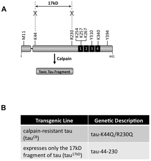
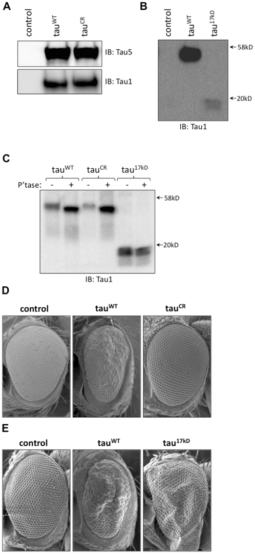
Alzheimer's disease and other related neurodegenerative disorders known as tauopathies are characterized by the accumulation of abnormally phosphorylated and aggregated forms of the microtubule-associated protein tau. Several laboratories have identified a 17 kD proteolytic fragment of tau in degenerating neurons and in numerous cell culture models that is generated by calpain cleavage and speculated to contribute to tau toxicity. In the current study, we employed a Drosophila tauopathy model to investigate the importance of calpain-mediated tau proteolysis in contributing to tau neurotoxicity in an animal model of human neurodegenerative disease. We found that mutations that disrupted endogenous calpainA or calpainB activity in transgenic flies suppressed tau toxicity. Expression of a calpain-resistant form of tau in Drosophila revealed that mutating the putative calpain cleavage sites that produce the 17 kD fragment was sufficient to abrogate tau toxicity in vivo. Furthermore, we found significant toxicity in the fly retina associated with expression of only the 17 kD tau fragment. Collectively, our data implicate calpain-mediated proteolysis of tau as an important pathway mediating tau neurotoxicity in vivo.


Conflict of interest statement

Competing Interests: The authors have declared that no competing interests exist.

Figures

Figure 1
Figure 1. Colocalization of tau and calpain in Drosophila neurons.
Neuronal cells cultured from late 3rd instar larvae expressing tauWT were stained for tau in red (A and D), calpain A and calpain B in green (B and E, respectively), with colocalization shown in yellow (C and F). The expression pattern of calpain A and B was similar to that of tau, with the primary site of colocalization in the neuronal cell body. Scale bar  = 5 µM.
Figure 2
Figure 2. Calpain mutants suppress tau-induced toxicity in the fly eye.
As shown using scanning electron microscopy, compared to the normal external appearance of control flies (w1118 in A; GMR-GAL4/+ in B), flies expressing wild-type human tau (tauWT; C) displayed the moderately rough eye phenotype characterisitc of tau toxicity. Genetic disruption of calpain A (D–F) or calpain B (G–I) using either deficiency lines (D and G) or P-element insertions (E–F and H–I) effectively suppressed tau toxicity.
Figure 3
Figure 3. Calpain protein expression level and enzyme activity are decreased in calpain mutants.
A. Using antibodies specific to calpain A or calpain B, western blot analysis showed that compared to tauWT-expressing flies, all of the calpain mutant lines have lower levels of calpain expression (top). Tau expression levels for all of the calpain mutants are equivalent to the expression level of tau in tauWT-expressing flies using both Tau 5 and Tau-1 antibodies (bottom two panels). B. When normalized to the level of calpain in tauWT-expressing flies, densitometric analysis revealed reduced calpain expression in all calpain mutant lines tested (top). The values shown represent the mean of three or more independent experiments. When normalized to calpain activity in tauWT-expressing flies, all calpA and calpB mutants have reduced calpain activity (bottom). The values shown represent the mean of enzyme activity measured in triplicate. Error bars for both graphs represent the standard error of the mean and data points indicated with asterisks are significant. *P<0.05, **P<0.01 (unpaired student t-test).
Figure 4
Figure 4. Cleavage of tau by calpain creates the 17 kD fragment.
A. Schematic of 0N4R tau with the nine putative calpain cleave sites indicated. Proteolysis at lysine 44 (K44) and arginine 230 (R230) yields a 17 kD fragment of tau. Abbreviations: methionine (M), lysine (K), arginine (R), and tyrosine (Y). B. The K44Q/R230Q mutation is predicted to disrupt the recognition motif for calpain at K44 and R230 and thus creates a calpain-resistant (tauCR) form of tau for expression in Drosophila. PCR was used to create a second tau mutant (tau17kD) in which only the amino acids corresponding to the reputed toxic 17 kD fragment (aa 44-230) are expressed in Drosophila.
Figure 5
Figure 5. Modifying two putative calpain cleavage sites in tau significantly impacts tau toxicity in vivo.
A. Western blot analysis of fly head homogenates revealed equivalent tau expression levels in tauCR-expressing flies compared to tauWT-expressing flies. There is no cross-reactivity of the Tau-1 or Tau 5 antibody with homogenates from control flies (GMR/+). B. Expression of the 17 kD fragment could not be detected at 25°C and instead required crosses to be conducted at 31°C. Possessing only the coding sequence for amino acids 44-230, tau17kD-expressing flies made only a ∼17 kD tau fragment that is found at lower abundance than that of full-length tau found in tauWT–expressing fly head homogenates with equivalent protein loading. C. Treatment of fly head homogenates from tauWT-, tauCR- and tau17kD-expressing flies with lambda phosphatase indicated that both the wild-type and calpain-resistant forms of tau experienced a mobility shift with phosphatase treatment whereas the 17 kD fragment was resistant to phosphatase treatment. To ensure we would visualize a potential molecular weight shift of the 17 kD fragment by western blot, the phosphatase assay employed three times more fly head homogenate from tau17kD-expressing flies than tauWT or tauCR flies. D. When compared to the rough eye phenotype in tauWT-expressing flies, tauCR flies demonstrated no detectable tau toxicity. E. At 31°C, transgenic flies that express only the 17 kD tau fragment (tau17kD) showed equivalent or slightly enhanced tau toxicity compared to the toxicity associated with expressing tauWT in the retina, suggesting that the 17 kD fragment possesses intrinsic toxicity in vivo. Control flies in D and E are GMR/+.

References

    1. Hong M, Zhukareva V, Vogelsberg-Ragaglia V, Wszolek Z, Reed L, et al. Mutation-specific functional impairments in distinct tau isoforms of hereditary FTDP-17. Science. 1998;282:1914–1917. - PubMed
    1. Poorkaj P, Bird TD, Wijsman E, Nemens E, Garruto RM, et al. Tau is a candidate gene for chromosome 17 frontotemporal dementia. Ann Neurol. 1998;43:815–825. - PubMed
    1. Hutton M, Lendon CL, Rizzu P, Baker M, Froelich S, et al. Association of missense and 5′-splice-site mutations in tau with the inherited dementia FTDP-17. Nature. 1998;393:702–705. - PubMed
    1. Spillantini MG, Murrell JR, Goedert M, Farlow MR, Klug A, et al. Mutation in the tau gene in familial multiple system tauopathy with presenile dementia. Proc Natl Acad Sci U S A. 1998;95:7737–7741. - PMC - PubMed
    1. Wittmann CW, Wszolek MF, Shulman JM, Salvaterra PM, Lewis J, et al. Tauopathy in Drosophila: neurodegeneration without neurofibrillary tangles. Science. 2001;293:711–714. - PubMed

Publication types

MeSH terms

LinkOut - more resources