

CD4+CD25+ regulatory cells contribute to the regulation of colonic Th2 granulomatous pathology caused by schistosome infection
- PMID: 21858239
- PMCID: PMC3153428
- DOI: 10.1371/journal.pntd.0001269
CD4+CD25+ regulatory cells contribute to the regulation of colonic Th2 granulomatous pathology caused by schistosome infection
Abstract
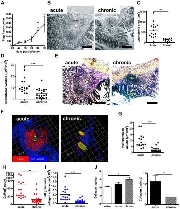
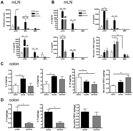
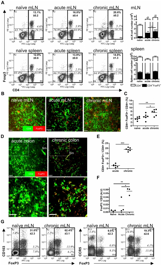
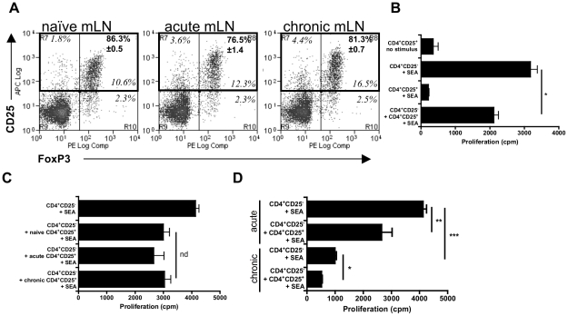
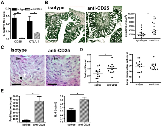
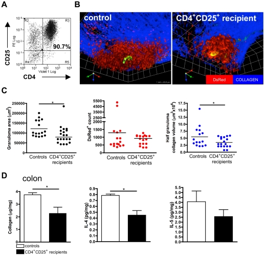
Eggs of the helminth Schistosoma mansoni accumulate in the colon following infection and generate Th2-biassed inflammatory granulomas which become down- modulated in size as the infection proceeds to chronicity. However, although CD4+CD25+FoxP3+ regulatory T cells (T(regs)) are known to suppress Th1-mediated colitis, it is not clear whether they control Th2-associated pathologies of the large intestine which characterise several helminth infections. Here we used a novel 3D-multiphoton confocal microscopy approach to visualise and quantify changes in the size and composition of colonic granulomas at the acute and chronic phases of S. mansoni infection. We observed decreased granuloma size, as well as reductions in the abundance of DsRed+ T cells and collagen deposition at 14 weeks (chronic) compared to 8 weeks (acute) post-infection. Th2 cytokine production (i.e. IL-4, IL-5) in the colonic tissue and draining mesenteric lymph node (mLN) decreased during the chronic phase of infection, whilst levels of TGF-β1 increased, co-incident with reduced mLN proliferative responses, granuloma size and fibrosis. The proportion of CD4+CD25+FoxP3+T(regs): CD4+ cells in the mLN increased during chronic disease, while within colonic granulomas there was an approximate 4-fold increase. The proportion of CD4+CD25+FoxP3+T(regs) in the mLN that were CD103+ and CCR5+ also increased indicating an enhanced potential to home to intestinal sites. CD4+CD25+ cells suppressed antigen-specific Th2 mLN cell proliferation in vitro, while their removal during chronic disease resulted in significantly larger granulomas, partial reversal of Th2 hypo-responsiveness and an increase in the number of eosinophils in colonic granulomas. Finally, transfer of schistosome infection-expanded CD4+CD25+T(regs) down-modulated the development of colonic granulomas, including collagen deposition. Therefore, CD4+CD25+FoxP3+T(regs) appear to control Th2 colonic granulomas during chronic infection, and are likely to play a role in containing pathology during intestinal schistosomiasis.
Conflict of interest statement
The authors have declared that no competing interests exist.
Figures
Similar articles
-
Retroviral Foxp3 gene transfer ameliorates liver granuloma pathology in Schistosoma mansoni infected mice.Immunology. 2005 Mar;114(3):410-7. doi: 10.1111/j.1365-2567.2004.02083.x. Immunology. 2005. PMID: 15720442 Free PMC article.
-
CCR8 is expressed by antigen-elicited, IL-10-producing CD4+CD25+ T cells, which regulate Th2-mediated granuloma formation in mice.J Immunol. 2005 Feb 15;174(4):1962-70. doi: 10.4049/jimmunol.174.4.1962. J Immunol. 2005. PMID: 15699124 Free PMC article.
-
Control of Schistosoma mansoni egg-induced inflammation by IL-4-responsive CD4(+)CD25(-)CD103(+)Foxp3(-) cells is IL-10-dependent.Eur J Immunol. 2010 Oct;40(10):2837-47. doi: 10.1002/eji.200940075. Eur J Immunol. 2010. PMID: 20821727
-
Experimental models of Schistosoma mansoni infection.Mem Inst Oswaldo Cruz. 2002 Oct;97(7):917-40. doi: 10.1590/s0074-02762002000700002. Mem Inst Oswaldo Cruz. 2002. PMID: 12471417 Review.
-
T Lymphocyte-Mediated Liver Immunopathology of Schistosomiasis.Front Immunol. 2020 Feb 18;11:61. doi: 10.3389/fimmu.2020.00061. eCollection 2020. Front Immunol. 2020. PMID: 32132991 Free PMC article. Review.
Cited by
-
Heat Shock Protein 60 in Eggs Specifically Induces Tregs and Reduces Liver Immunopathology in Mice with Schistosomiasis Japonica.PLoS One. 2015 Sep 29;10(9):e0139133. doi: 10.1371/journal.pone.0139133. eCollection 2015. PLoS One. 2015. PMID: 26418003 Free PMC article.
-
Schistosome Egg Migration: Mechanisms, Pathogenesis and Host Immune Responses.Front Immunol. 2018 Dec 20;9:3042. doi: 10.3389/fimmu.2018.03042. eCollection 2018. Front Immunol. 2018. PMID: 30619372 Free PMC article. Review.
-
Schistosoma mansoni Larvae Do Not Expand or Activate Foxp3+ Regulatory T Cells during Their Migratory Phase.Infect Immun. 2015 Oct;83(10):3881-9. doi: 10.1128/IAI.00408-15. Epub 2015 Jul 20. Infect Immun. 2015. PMID: 26195548 Free PMC article.
-
HGF Gene Modification in Mesenchymal Stem Cells Reduces Radiation-Induced Intestinal Injury by Modulating Immunity.PLoS One. 2015 May 1;10(5):e0124420. doi: 10.1371/journal.pone.0124420. eCollection 2015. PLoS One. 2015. PMID: 25933295 Free PMC article.
-
FOXP3+ Regulatory T Cells in Hepatic Fibrosis and Splenomegaly Caused by Schistosoma japonicum: The Spleen May Be a Major Source of Tregs in Subjects with Splenomegaly.PLoS Negl Trop Dis. 2016 Jan 5;10(1):e0004306. doi: 10.1371/journal.pntd.0004306. eCollection 2016 Jan. PLoS Negl Trop Dis. 2016. PMID: 26731721 Free PMC article.
References
-
- Chitsulo L, Loverde P, Engels D. Schistosomiasis. Nat Rev Microbiol. 2004;2:12–13. - PubMed
-
- Steinmann P, Keiser J, Bos R, Tanner M, Utzinger J. Schistosomiasis and water resources development: systematic review, meta-analysis, and estimates of people at risk. Lancet Infect Dis. 2006;6:411–425. - PubMed
-
- Gryseels B, Polman K, Clerinx J, Kestens L. Human schistosomiasis. Lancet. 2006;368:1106–1118. - PubMed
-
- King CL. Initiation and regulation of disease in schistosomiasis. In: Mahmoud A, editor. Schistosomiasis. London: Imperial College Press; 2001. pp. 213–264.
Publication types
MeSH terms
Substances
Grants and funding
LinkOut - more resources
Full Text Sources
Research Materials
