. 2011 Sep;345(3):391-403.
doi: 10.1007/s00441-011-1229-z. Epub 2011 Aug 20.

Telocytes and putative stem cells in the lungs: electron microscopy, electron tomography and laser scanning microscopy

Affiliations

Telocytes and putative stem cells in the lungs: electron microscopy, electron tomography and laser scanning microscopy

Laurentiu M Popescu et al. Cell Tissue Res. 2011 Sep.

Abstract

This study describes a novel type of interstitial (stromal) cell - telocytes (TCs) - in the human and mouse respiratory tree (terminal and respiratory bronchioles, as well as alveolar ducts). TCs have recently been described in pleura, epicardium, myocardium, endocardium, intestine, uterus, pancreas, mammary gland, etc. (see www.telocytes.com ). TCs are cells with specific prolongations called telopodes (Tp), frequently two to three per cell. Tp are very long prolongations (tens up to hundreds of μm) built of alternating thin segments known as podomers (≤ 200 nm, below the resolving power of light microscope) and dilated segments called podoms, which accommodate mitochondria, rough endoplasmic reticulum and caveolae. Tp ramify dichotomously, making a 3-dimensional network with complex homo- and heterocellular junctions. Confocal microscopy reveals that TCs are c-kit- and CD34-positive. Tp release shed vesicles or exosomes, sending macromolecular signals to neighboring cells and eventually modifying their transcriptional activity. At bronchoalveolar junctions, TCs have been observed in close association with putative stem cells (SCs) in the subepithelial stroma. SCs are recognized by their ultrastructure and Sca-1 positivity. Tp surround SCs, forming complex TC-SC niches (TC-SCNs). Electron tomography allows the identification of bridging nanostructures, which connect Tp with SCs. In conclusion, this study shows the presence of TCs in lungs and identifies a TC-SC tandem in subepithelial niches of the bronchiolar tree. In TC-SCNs, the synergy of TCs and SCs may be based on nanocontacts and shed vesicles.


Figures
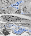
Fig. 1
Fig. 1
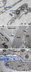
Transmission electron microscopy: telocytes (TCs) in mouse terminal bronchioles (a, b) and human lung (c). a One telocyte (blue) extends two telopodes (Tp1, Tp2) along the basement membrane of the bronchiolar epithelium. A third telopode (Tp3) is located among smooth muscle cells (SMC). Schwann cells and nerve endings appear in close vicinity to the telocyte. Characteristic dilated podoms (p) alternate with podomers (pm), thin segments along the Tp. b A peri-bronchiolar fibroblast is shown for comparison with TCs: note the numerous dilated cisternae of rough endoplasmic reticulum (rER) and mitochondria (m). The peri-bronchiolar interstitial space also has numerous collagen fibrils (coll) and vascular smooth muscle cells (vSMC). c TC and Tp border the epithelium at the level of the respiratory bronchiole. Pn I, type I pneumocyte; Pn II, type II pneumocyte. Scale bars 5 μm (a, c), 2 μm (b)
Fig. 2
Fig. 2
Transmission electron microscopy of telopodes (Tp) in the interstitium of mouse terminal bronchioles. a Note the characteristic thin podomeres (50–200 nm; below the resolving power of light microscope) (Tp1), as well as a specific podom (Tp2), a dilation containing mitochondria (m), the endoplasmic reticulum (ER) and caveolae (arrowheads). b Extremely thin telopodes (Tp1, Tp2; < 50 nm) can be seen bordering elastin fibers (elast) on both sides, adjacent to the bronchiolar epithelium and smooth muscle cells (SMC). Ci, ciliated cell. c A telopode (Tp) has two contacts (encircled), one with the basement membrane of the epithelium and another with the basal lamina of a smooth muscle cell (SMC). Note the dichotomic pattern of Tp branching. Scale bars 1 μm (ac)
Fig. 3
Fig. 3
Connections between telocytes. a,b Telopodes (Tp1, Tp2, Tp3) bordering the alveolar epithelium are connected by puncta adhaerentia junctions (white arrows). Dense structures (attachment plaques) connect Tp to the extracellular matrix (black arrows). Pn I, type I pneumocyte. c 60-nm sections show Tp (Tp1, Tp2) connected by manubria adhaerentia (dotted circles). d Exosomes containing shed vesicles (sv) emerge from the Tp. One telopode (Tp1) enfolds a large vesicle (lv) located in the extracellular space. Note the thickness of telopodes (Tp1, Tp2), which is comparable with the thickness of collagen fibrils (coll): approximately 50 nm (far below the resolving power of light microscope). Scale bars 1 μm (ad)
Fig. 4
Fig. 4
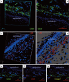
Confocal microscopy of thick sections (60 μm) of lung tissue from an adult mouse, with CD34 immunolabeling visible in the peribronchiolar space. a Volume reconstruction. b Confocal laser scanning microscopy. c c-kit immunolabeling, original volume reconstruction. d The same volume after deconvolution. Double immunofluorescence shows the labeling pattern for CD34 (e, green) and c-kit (f, red); the superimposed images are presented in g. Nuclei were counterstained with DAPI. Confocal images were collected using the 60× 1.2-NA water objective (z-axis step 0.16 μm)
Fig. 5
Fig. 5
Immunofluorescent labeling of Sca-1 positive cells (green) from the peri-bronchiolar space in the adult mouse lung, detected by confocal laser scanning microscopy. The nuclei were counterstained with DAPI (blue)
Fig. 6
Fig. 6
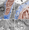
Telocytes and a possible stem cell niche at the level of the bronchoalveolar junction (mouse). a A group of putative stem cells (SC), underneath the alveolar epithelial cells (Pn I, type I pneumocytes; Pn II, type II pneumocytes). The cluster of SCs (brown) is surrounded by telopodes (Tp1, Tp2; blue). TC, telocyte; vSMC, vascular smooth muscle cells. b Higher magnification of the upper boxed area shows one telopode (Tp1, blue) in contact with a type I pneumocyte (Pn I, black arrow) and a brown-colored SC (white arrows). A shed vesicle visible in violet (asterisk). c Higher magnification of the lower boxed area in a shows a break in the basement membrane (bm). A telopode (Tp2) makes contact (arrow) with a type I pneumocyte (Pn I). d TEM image shows a contact point (arrow) between a telopode (Tp1) and a type I pneumocyte (Pn I). Scale bars 2 μm (a); 1 μm (bd)
Fig. 7
Fig. 7
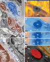
Transmission electron microscopy of mouse broncho-alveolar junction. a A telocyte (TC, blue) with numerous, abrupt emerging telopodes (Tp). Some Tp surround an array of putative stem cells (SC). vSMC, vascular smooth muscle cells; Macro, macrophage. b Higher magnification of the black boxed area in a shows that a Tp passes between putative stem cells (SC) and a macrophage (Macro). Small dense structures (white arrows) connect the telopode (Tp1) with SCs, while the larger dense structures, puncta adhaerentia (black arrows), connect Tp with the macrophage. A small shed vesicle (violet, asterisk) is visible between the Tp and SC. cg Electron tomography images of the red-boxed area in panel a. Digital sections from the grey area in panel c are enlarged in panel d. e, f Serial digital sections from lateral views of the tomographic volume at different levels. Images df show small bridging nanostructures (white arrows), which connect a Tp with an SC. A punctum adhaerens (black arrow) connects two Tp (Tp1, Tp2). dg, dense granule. g Three-dimensional representation of connecting nanostructures (green dots) between Tp and SC in the vicinity of a dense granule (red) from telopode Tp1. Scale bars 5 μm (a); 1 μm (b); 0.5 μm (c,d); 0.2 μm (eg)
Fig. 8
Fig. 8
a Electron tomography shows telopodes (Tp1, Tp2) with irregular contours. sv, shed vesicle. See also the 3D volume in Fig. S1. b A lateral view of the tomographic volume at the level of the double-ended arrow in a demonstrates that Tp2 is entirely embedded (arrows) in the thickness (250 nm) of the section. c Higher magnification of a digital section through the bright area in reveals that caveolae present in Tp1 establish contacts with the cellular membrane on the junctional side. Telopodes are connected by a cluster of contact points (arrows). Tp2 is extremely narrow (< 50 nm) at the level of the junction. c, caveolae. d A lateral view of the tomographic volume at the level of one caveola (c) shows a 10-nm contact point (arrow) between telopodes (Tp1,Tp2). Scale bars 0.5 μm (a); 0.2 μm (bd)
Fig. 9
Fig. 9
Immunohistochemical reaction for VEGF in the adult mouse lung. VEGF expression in telocytes (TCs) is more evident around bronchioles (a), as well as between a bronchiole and the accompanying vein (b). Tp extend in opposite directions and interconnect with similar neighboring cells. Nuclei were counterstained with hematoxylin. Scale bars 5 μm

Similar articles

Cited by

References

    1. Bani D, Formigli L, Gherghiceanu M, Faussone-Pellegrini MS. Telocytes as supporting cells for myocardial tissue organization in developing and adult heart. J Cell Mol Med. 2010;14:2531–2538. doi: 10.1111/j.1582-4934.2010.01119.x. - DOI - PMC - PubMed
    1. Barth M, Schumacher H, Kuhn C, Akhyari P, Lichtenberg A, Franke WW. Cordial connections: molecular ensembles and structures of adhering junctions connecting interstitial cells of cardiac valves in situ and in cell culture. Cell Tissue Res. 2009;337:63–77. doi: 10.1007/s00441-009-0806-x. - DOI - PubMed
    1. Bastiani M, Parton RG. Caveolae at a glance. J Cell Sci. 2010;123:3831–3836. doi: 10.1242/jcs.070102. - DOI - PubMed
    1. Bell G, Mooers AO. Size and complexity among multicellular organisms. Biol J Linn Soc Lond. 1997;60:345–363. doi: 10.1111/j.1095-8312.1997.tb01500.x. - DOI
    1. Bojin FM, Gavriliuc OI, Cristea MI, Tanasie G, Tatu CS, Panaitescu C, Paunescu V (2011) Telocytes within human skeletal muscle stem cell niche. J Cell Mol Med. doi:10.1111/j.1582-4934.2011.01386.x - PMC - PubMed

Publication types