Cerenkov emission induced by external beam radiation stimulates molecular fluorescence
- PMID: 21859013
- PMCID: PMC3139507
- DOI: 10.1118/1.3592646
Cerenkov emission induced by external beam radiation stimulates molecular fluorescence
Abstract
Purpose: Cerenkov emission is induced when a charged particle moves faster than the speed of light in a given medium. Both x-ray photons and electrons produce optical Cerenkov photons in everyday radiation therapy of tissue; yet, this phenomenon has never been fully documented. This study quantifies the emissions and also demonstrates that the Cerenkov emission can excite a fluorophore, protoporphyrin IX (PpIX), embedded in biological phantoms.
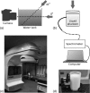
Methods: In this study, Cerenkov emission induced by radiation from a clinical linear accelerator is investigated. Biological mimicking phantoms were irradiated with x-ray photons, with energies of 6 or 18 MV, or electrons at energies 6, 9, 12, 15, or 18 MeV. The Cerenkov emission and the induced molecular fluorescence were detected by a camera or a spectrometer equipped with a fiber optic cable.
Results: It is shown that both x-ray photons and electrons, at MeV energies, produce optical Cerenkov photons in tissue mimicking media. Furthermore, we demonstrate that the Cerenkov emission can excite a fluorophore, protoporphyrin IX (PpIX), embedded in biological phantoms.
Conclusions: The results here indicate that molecular fluorescence monitoring during external beam radiotherapy is possible.
Figures


Similar articles
-
Superficial dosimetry imaging based on Čerenkov emission for external beam radiotherapy with megavoltage x-ray beam.Med Phys. 2013 Oct;40(10):101914. doi: 10.1118/1.4821543. Med Phys. 2013. PMID: 24089916 Free PMC article.
-
Application of Cerenkov radiation generated in plastic optical fibers for therapeutic photon beam dosimetry.J Biomed Opt. 2013 Feb;18(2):27001. doi: 10.1117/1.JBO.18.2.027001. J Biomed Opt. 2013. PMID: 23377008
-
Čerenkov excited fluorescence tomography using external beam radiation.Opt Lett. 2013 Apr 15;38(8):1364-6. doi: 10.1364/OL.38.001364. Opt Lett. 2013. PMID: 23595486 Free PMC article.
-
Optical Imaging of Ionizing Radiation from Clinical Sources.J Nucl Med. 2016 Nov;57(11):1661-1666. doi: 10.2967/jnumed.116.178624. Epub 2016 Sep 29. J Nucl Med. 2016. PMID: 27688469 Free PMC article. Review.
-
Cerenkov radiation-activated probes for deep cancer theranostics: a review.Theranostics. 2022 Oct 24;12(17):7404-7419. doi: 10.7150/thno.75279. eCollection 2022. Theranostics. 2022. PMID: 36438500 Free PMC article. Review.
Cited by
-
Projection imaging of photon beams by the Čerenkov effect.Med Phys. 2013 Jan;40(1):012101. doi: 10.1118/1.4770286. Med Phys. 2013. PMID: 23298103 Free PMC article.
-
Effects of core titanium crystal dimension and crystal phase on ROS generation and tumour accumulation of transferrin coated titanium dioxide nanoaggregates.RSC Adv. 2020;10(40):23759-23766. doi: 10.1039/d0ra01878c. Epub 2020 Jun 23. RSC Adv. 2020. PMID: 32774845 Free PMC article.
-
Activatable probes based on distance-dependent luminescence associated with Cerenkov radiation.Angew Chem Int Ed Engl. 2013 Jul 22;52(30):7756-60. doi: 10.1002/anie.201302564. Epub 2013 Jun 13. Angew Chem Int Ed Engl. 2013. PMID: 23765506 Free PMC article.
-
Time-gated Cherenkov emission spectroscopy from linear accelerator irradiation of tissue phantoms.Opt Lett. 2012 Apr 1;37(7):1193-5. doi: 10.1364/OL.37.001193. Opt Lett. 2012. PMID: 22466192 Free PMC article.
-
Radioluminescent nanophosphors enable multiplexed small-animal imaging.Opt Express. 2012 May 21;20(11):11598-604. doi: 10.1364/OE.20.011598. Opt Express. 2012. PMID: 22714145 Free PMC article.
References
-
- Cerenkov P. A., “Visible emission of clean liquids by action of γ radiation,” Dok. Akad. Nauk SSSR 2, 451–454 (1934).
-
- Ross H. H., “Measurement of β-emitting nuclides using Cerenkov radiation,” Anal. Chem. 41(10), 1260–1265 (1969).10.1021/ac60279a011 - DOI
Publication types
MeSH terms
Grants and funding
LinkOut - more resources
Full Text Sources
Other Literature Sources

