

A quality assurance protocol for diffusion tensor imaging using the head phantom from American College of Radiology
- PMID: 21859042
- PMCID: PMC3145225
- DOI: 10.1118/1.3595111
A quality assurance protocol for diffusion tensor imaging using the head phantom from American College of Radiology
Abstract
Purpose: To propose a quality assurance procedure for routine clinical diffusion tensor imaging (DTI) using the widely available American College of Radiology (ACR) head phantom.
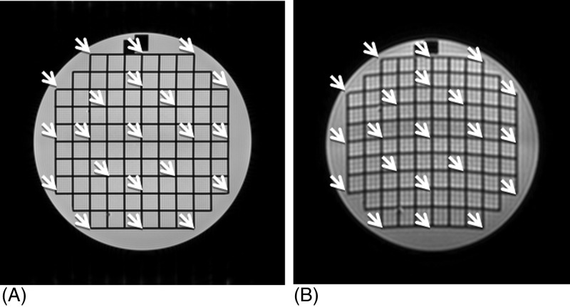
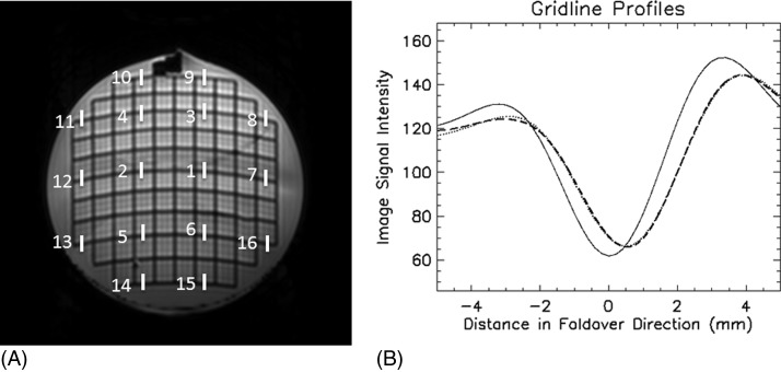
Methods: Analysis was performed on the data acquired at 1.5 and 3.0 T on whole body clinical MRI scanners using the ACR phantom and included the following: (1) the signal-to-noise ratio (SNR) at the center and periphery of the phantom, (2) image distortion by EPI readout relative to spin echo imaging, (3) distortion of high-b images relative to the b= 0 image caused by diffusion encoding, and (4) determination of fractional anisotropy (FA) and mean diffusivity (MD) measured with region-of-interest (ROI) and pixel-based approaches. Reproducibility of the measurements was assessed by five repetitions of data acquisition on each scanner.
Results: The SNR at the phantom center was approximately half of that near the periphery at both 1.5 and 3 T. The image distortion by the EPI readout was up to 7 mm at 1.5 T and 10 mm at 3 T. The typical distortion caused by eddy currents from diffusion encoding was on the order of 0.5 mm. The difference between ROI-based and pixel-based MD quantification was 1.4% at 1.5 T and 0.3% at 3 T. The ROI-based MD values were in close agreement (within 2%) with the reference values. The ROI-based FA values were approximately a factor of 10 smaller than pixel-based values and less than 0.01. The measurement reproducibility was sufficient for quality assurance (QA) purposes.
Conclusions: This QA approach is simple to perform and evaluates key aspects of the scanner performance for DTI data acquisition using a widely available phantom.
Figures

References
Publication types
MeSH terms
LinkOut - more resources
Full Text Sources
