

Rapid loss of bone mass and strength in mice after abdominal irradiation
- PMID: 21859327
- PMCID: PMC3209530
- DOI: 10.1667/rr2505.1
Rapid loss of bone mass and strength in mice after abdominal irradiation
Abstract
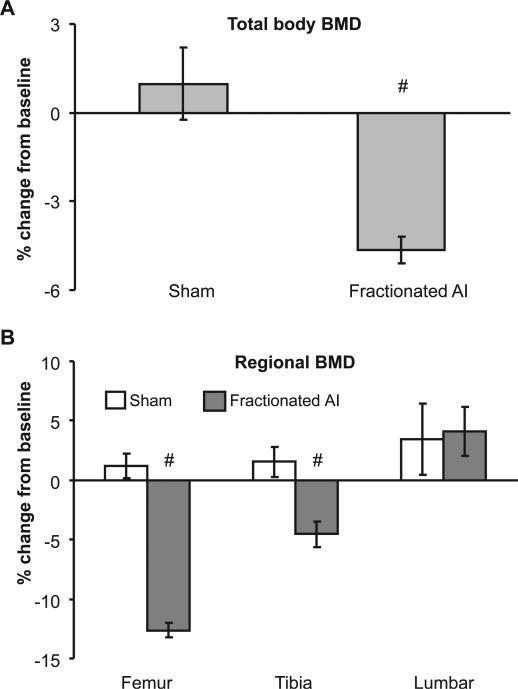
Localized irradiation is a common treatment modality for malignancies in the pelvic-abdominal cavity. We report here on the changes in bone mass and strength in mice 7-14 days after abdominal irradiation. Male C57BL/6 mice of 10-12 weeks of age were given a single-dose (0, 5, 10, 15 or 20 Gy) or fractionated (3 Gy × 2 per day × 7.5 days) X rays to the abdomen and monitored daily for up to 14 days. A decrease in the serum bone formation marker and ex vivo osteoblast differentiation was detected 7 days after a single dose of radiation, with little change in the serum bone resorption marker and ex vivo osteoclast formation. A single dose of radiation elicited a loss of bone mineral density (BMD) within 14 days of irradiation. The BMD loss was up to 4.1% in the whole skeleton, 7.3% in tibia, and 7.7% in the femur. Fractionated abdominal irradiation induced similar extents of BMD loss 10 days after the last fraction: 6.2% in the whole skeleton, 5.1% in tibia, and 13.8% in the femur. The loss of BMD was dependent on radiation dose and was more profound in the trabecula-rich regions of the long bones. Moreover, BMD loss in the total skeleton and the femurs progressed with time. Peak load and stiffness in the mid-shaft tibia from irradiated mice were 11.2-14.2% and 11.5-25.0% lower, respectively, than sham controls tested 7 days after a single-dose abdominal irradiation. Our data demonstrate that abdominal irradiation induces a rapid loss of BMD in the mouse skeleton. These effects are bone type- and region-specific but are independent of radiation fractionation. The radiation-induced abscopal damage to the skeleton is manifested by the deterioration of biomechanical properties of the affected bone.
Figures


Similar articles
-
Total-body irradiation of postpubertal mice with (137)Cs acutely compromises the microarchitecture of cancellous bone and increases osteoclasts.Radiat Res. 2009 Mar;171(3):283-9. doi: 10.1667/RR1463.1. Radiat Res. 2009. PMID: 19267555
-
A Mouse Model for Skeletal Structure and Function Changes Caused by Radiation Therapy and Estrogen Deficiency.Calcif Tissue Int. 2020 Feb;106(2):180-193. doi: 10.1007/s00223-019-00617-x. Epub 2019 Oct 3. Calcif Tissue Int. 2020. PMID: 31583426
-
Single-Limb Irradiation Induces Local and Systemic Bone Loss in a Murine Model.J Bone Miner Res. 2015 Jul;30(7):1268-79. doi: 10.1002/jbmr.2458. Epub 2015 Jun 8. J Bone Miner Res. 2015. PMID: 25588731 Free PMC article.
-
Effects of Radiotherapy Upon Bone Structure-Strength Relationships Vary With Sex and Fractionation of Dosing.Iowa Orthop J. 2023;43(1):77-86. Iowa Orthop J. 2023. PMID: 37383848 Free PMC article.
-
Lowering iron level protects against bone loss in focally irradiated and contralateral femurs through distinct mechanisms.Bone. 2019 Mar;120:50-60. doi: 10.1016/j.bone.2018.10.005. Epub 2018 Oct 7. Bone. 2019. PMID: 30304704
Cited by
-
Suppression of Sclerostin Alleviates Radiation-Induced Bone Loss by Protecting Bone-Forming Cells and Their Progenitors Through Distinct Mechanisms.J Bone Miner Res. 2017 Feb;32(2):360-372. doi: 10.1002/jbmr.2996. Epub 2016 Oct 20. J Bone Miner Res. 2017. PMID: 27635523 Free PMC article.
-
Melatonin can Ameliorate Radiation-Induced Oxidative Stress and Inflammation-Related Deterioration of Bone Quality in Rat Femur.Inflammation. 2016 Jun;39(3):1134-40. doi: 10.1007/s10753-016-0347-x. Inflammation. 2016. PMID: 27052631
-
Total body irradiation is associated with long-term deficits in femoral bone structure but not mechanical properties in male rhesus macaques.Sci Rep. 2024 Oct 8;14(1):23379. doi: 10.1038/s41598-024-75363-8. Sci Rep. 2024. PMID: 39379502 Free PMC article.
-
Irradiation alters the differentiation potential of bone marrow mesenchymal stem cells.Mol Med Rep. 2016 Jan;13(1):213-23. doi: 10.3892/mmr.2015.4539. Epub 2015 Nov 10. Mol Med Rep. 2016. PMID: 26572960 Free PMC article.
-
Ionizing radiation causes active degradation and reduces matrix synthesis in articular cartilage.Int J Radiat Biol. 2013 Apr;89(4):268-77. doi: 10.3109/09553002.2013.747015. Epub 2012 Dec 12. Int J Radiat Biol. 2013. PMID: 23134087 Free PMC article.
References
-
- Orton CG. Uses of therapeutic x rays in medicine. Health Phys. 1995;69:662–76. - PubMed
-
- Bernier J, Hall EJ, Giaccia A. Radiation oncology: a century of achievements. Nat Rev Cancer. 2004;4:737–47. - PubMed
-
- Cohen L, Schultheiss TE, Hendrickson FR, Mansell J, Saroja KR, Lennox A. Normal tissue reactions and complications following high-energy neutron beam therapy: I. Crude response rates. Int J Radiat Oncol Biol Phys. 1989;16:73–8. - PubMed
-
- Stone HB, Coleman CN, Anscher MS, McBride WH. Effects of radiation on normal tissue: consequences and mechanisms. Lancet Oncol. 2003;4:529–36. - PubMed
-
- Smith RP, Heron DE, Huq MS, Yue NJ. Modern radiation treatment planning and delivery—from Röntgen to real time. Hematol Oncol Clin N Am. 2006;20:45–62. - PubMed
Publication types
MeSH terms
Grants and funding
LinkOut - more resources
Full Text Sources
Medical
