Review
. 2011 Nov;31(11):803-12.
doi: 10.1089/jir.2011.0045. Epub 2011 Aug 22.

Type I interferon and systemic lupus erythematosus

Affiliations
Review

Type I interferon and systemic lupus erythematosus

Keith B Elkon et al. J Interferon Cytokine Res. 2011 Nov.

Abstract

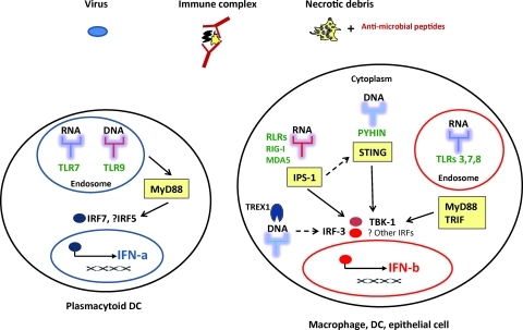
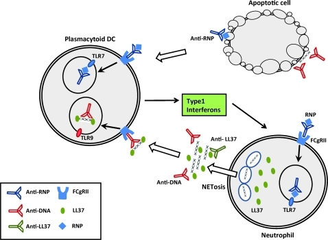
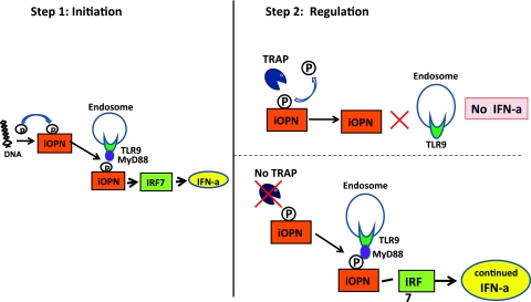
Systemic lupus erythematosus (SLE) is a complex systemic autoimmune disease associated with multiple immunologic abnormalities. Prominent among these is upregulation of type I interferon (IFN)?a powerful immune adjuvant. IFN is, in part, produced in SLE in response to autoantigens in the form of self-nucleic acids and their associated nuclear proteins. Sources of these autoantigens include apoptotic and necrotic cells as well as neutrophils undergoing a specific form of cell death called NETosis. Although plasmacytoid dendritic cells are the main producers of IFN-a, other cells are important regulators of this process. Both genetic and environmental risk factors play a role in the development and pathogenesis of SLE. Further highlighting the importance of IFN, candidate gene and genome-wide association studies have identified a number of genes involved in type I IFN pathways associated with SLE. In this review, 3 monogenic deficiencies that result in lupus-like phenotypes and several polygenic variants that have been consistently associated with SLE are highlighted, and the relationship of these genes to IFN-a production is discussed. Clinical associations of the type I IFN pathway and the use of IFN-blocking agents as therapeutic agents in SLE are also reviewed.


Figures

FIG. 1.
FIG. 1.
Innate signaling pathways leading to type I interferon (IFN) production. Various triggers—viruses, nucleic acid-containing immune complexes, and necrotic debris associated with antimicrobial peptides—are sensed by pattern recognition receptors in cells, including endosomal Toll-like receptors (TLRs) and cytoplasmic RIG-I-like receptors (RLRs) and pyrin and HIN200 domain-containing protein (PYHIN) receptors and lead to IFN production. TREX1 negatively regulates interferon stimulation by cytoplasmic DNA. Yellow boxes represent adapter molecules necessary for downstream signal transduction. IPS-1, interferon-beta promotor stimulator 1; IRF, interferon regulatory factor; MyD88, myeloid differentiation factor 88; STING, stimulator of interferon gene; TRIF, Toll-receptor associated activator of interferon; TBK-1, TANK binding kinase1.
FIG. 2.
FIG. 2.
Stimulation of type I interferon by cell debris and immune complexes. Immune complexes (ICs) containing ribonucleoproteins (RNP) can be generated by the binding of autoantibodies to RNP containing antigens released by apoptotic or necrotic cells (top right). In addition, ICs containing deoxyribonucleoproteins (DNP) can be generated by the binding of autoantibodies to DNA or antimicrobial peptides, such as LL37, after NETosis of neutrophils (bottom right). Ultimately, these ICs are internalized by FcgR on plasmacytoid dendritic cells (pDC) and activate TLR7 or 9 resulting in the production of type I interferon in systemic lupus erythematosus (SLE; top left).
FIG. 3.
FIG. 3.
Hypothetical 2-step model depicting how tartrate-resistant acid phosphatase (TRAP) deficiency may regulate IFN-a production in pDCs. In step 1, the presence of cytoplasmic DNA leads to phosphorylation of intracellular osteopontin (iOPN). This in turn facilitates binding to MyD88/TLR9, activation of IRF7, and IFN-a production. In step 2, in normal cells (top right) TRAP dephosphorylates iOPN leading to reduction in IFN-a production (negative regulation). In the absence of TRAP (bottom right), iOPN remains phosphorylated, resulting in persistent activation of the TLR9/MyD88 pathway and continued IFN-a production.

References

    1. Andersson G. Ek-Rylander B. Hollberg K. Ljusberg-Sjolander J. Lang P. Norgard M. Wang Y. Zhang SJ. TRACP as an osteopontin phosphatase. J Bone Miner Res. 2003;18(10):1912–1915. - PubMed
    1. Baccala R. Hoebe K. Kono DH. Beutler B. Theofilopoulos AN. TLR-dependent and TLR-independent pathways of type I interferon induction in systemic autoimmunity. Nat Med. 2007;13(5):543–551. - PubMed
    1. Baechler EC. Batliwalla FM. Karypis G. Gaffney PM. Ortmann WA. Espe KJ. Shark KB. Grande WJ. Hughes KM. Kapur V. Interferon-inducible gene expression signature in peripheral blood cells of patients with severe lupus. Proc Natl Acad Sci U S A. 2003;100(5):2610–2615. others. - PMC - PubMed
    1. Barber GN. Innate immune DNA sensing pathways: STING, AIMII and the regulation of interferon production and inflammatory responses. Curr Opin Immunol. 2011;23(1):10–20. - PMC - PubMed
    1. Bauer JW. Petri M. Batliwalla FM. Koeuth T. Wilson J. Slattery C. Panoskaltsis-Mortari A. Gregersen PK. Behrens TW. Baechler EC. Interferon-regulated chemokines as biomarkers of systemic lupus erythematosus disease activity: a validation study. Arthritis Rheum. 2009;60(10):3098–3107. - PMC - PubMed

Publication types

MeSH terms

Substances