

Magnetic resonance imaging detects significant sex differences in human myocardial strain
- PMID: 21859466
- PMCID: PMC3180436
- DOI: 10.1186/1475-925X-10-76
Magnetic resonance imaging detects significant sex differences in human myocardial strain
Abstract
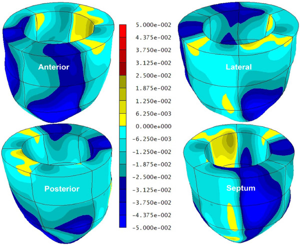
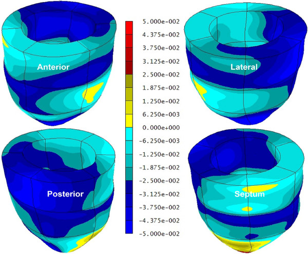
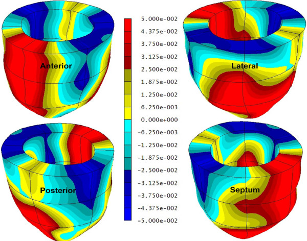
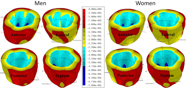
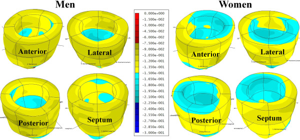
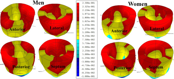
Background: The pathophysiology responsible for the significant outcome disparities between men and women with cardiac disease is largely unknown. Further investigation into basic cardiac physiological differences between the sexes is needed. This study utilized magnetic resonance imaging (MRI)-based multiparametric strain analysis to search for sex-based differences in regional myocardial contractile function.
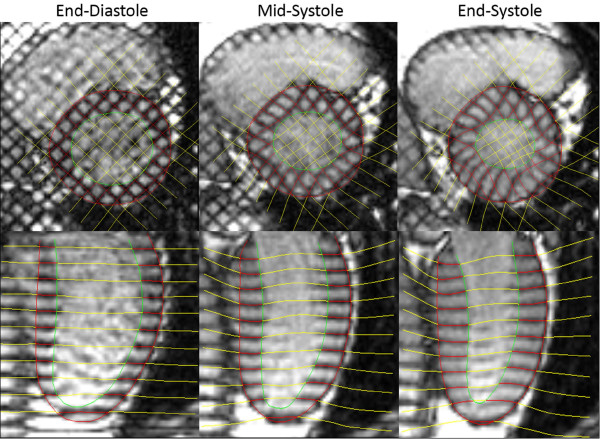
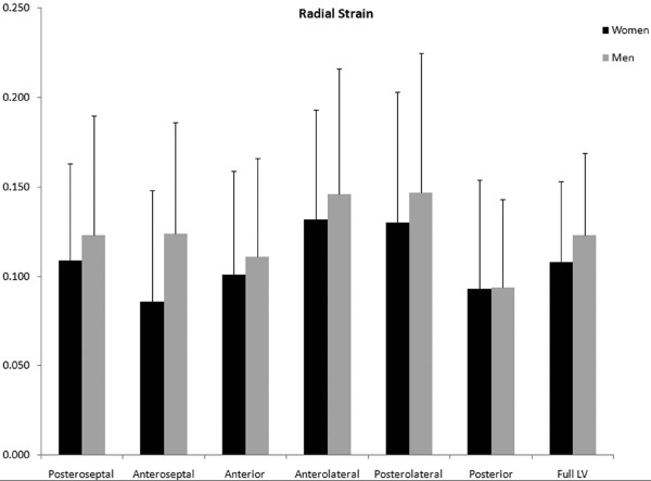
Methods: End-systolic strain (circumferential, longitudinal, and radial) was interpolated from MRI-based radiofrequency tissue tagging grid point displacements in each of 60 normal adult volunteers (32 females).
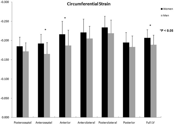
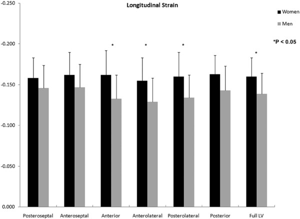
Results: The average global left ventricular (LV) strain among normal female volunteers (n = 32) was significantly larger in absolute value (functionally better) than in normal male volunteers (n = 28) in both the circumferential direction (Male/Female = -0.19 ± 0.02 vs. -0.21 ± 0.02; p = 0.025) and longitudinal direction (Male/Female = -0.14 ± 0.03 vs. -0.16 ± 0.02; p = 0.007).
Conclusions: The finding of significantly larger circumferential and longitudinal LV strain among normal female volunteers suggests that baseline contractile differences between the sexes may contribute to the well-recognized divergence in cardiovascular disease outcomes. Further work is needed in order to determine the pathologic changes that occur in LV strain between women and men with the onset of cardiovascular disease.
Figures
Similar articles
-
Myocardial viability mapping by magnetic resonance-based multiparametric systolic strain analysis.Ann Thorac Surg. 2008 Nov;86(5):1546-53. doi: 10.1016/j.athoracsur.2008.06.072. Ann Thorac Surg. 2008. PMID: 19049746 Free PMC article.
-
Magnetic resonance imaging-based multiparametric systolic strain analysis and regional contractile heterogeneity in patients with dilated cardiomyopathy.J Heart Lung Transplant. 2009 Apr;28(4):388-94. doi: 10.1016/j.healun.2008.12.018. Epub 2009 Feb 13. J Heart Lung Transplant. 2009. PMID: 19332267 Free PMC article.
-
Left ventricular global myocardial strain assessment comparing the reproducibility of four commercially available CMR-feature tracking algorithms.Eur Radiol. 2018 Dec;28(12):5137-5147. doi: 10.1007/s00330-018-5538-4. Epub 2018 Jun 5. Eur Radiol. 2018. PMID: 29872912
-
The functional role of longitudinal, circumferential, and radial myocardial deformation for regulating the early impairment of left ventricular contraction and relaxation in patients with cardiovascular risk factors: a study with two-dimensional strain imaging.J Am Soc Echocardiogr. 2008 Oct;21(10):1138-44. doi: 10.1016/j.echo.2008.07.016. J Am Soc Echocardiogr. 2008. PMID: 18926389
-
MRI-Derived Myocardial Strain Measures in Normal Subjects.JACC Cardiovasc Imaging. 2018 Feb;11(2 Pt 1):196-205. doi: 10.1016/j.jcmg.2016.12.025. Epub 2017 May 17. JACC Cardiovasc Imaging. 2018. PMID: 28528164
Cited by
-
Relationship Between Left Ventricular Structural and Metabolic Remodeling in Type 2 Diabetes.Diabetes. 2016 Jan;65(1):44-52. doi: 10.2337/db15-0627. Epub 2015 Oct 5. Diabetes. 2016. PMID: 26438611 Free PMC article.
-
Ectopic and Visceral Fat Deposition in Lean and Obese Patients With Type 2 Diabetes.J Am Coll Cardiol. 2016 Jul 5;68(1):53-63. doi: 10.1016/j.jacc.2016.03.597. J Am Coll Cardiol. 2016. PMID: 27364051 Free PMC article.
-
Reference ranges ("normal values") for cardiovascular magnetic resonance (CMR) in adults and children: 2020 update.J Cardiovasc Magn Reson. 2020 Dec 14;22(1):87. doi: 10.1186/s12968-020-00683-3. J Cardiovasc Magn Reson. 2020. PMID: 33308262 Free PMC article. Review.
-
Sex Differences in Cardiac Flow Dynamics of Healthy Volunteers.Radiol Cardiothorac Imaging. 2020 Feb;2(1):e190058. doi: 10.1148/ryct.2020190058. Epub 2020 Feb 27. Radiol Cardiothorac Imaging. 2020. PMID: 32666051 Free PMC article.
-
Sex differences in the evolution of left ventricle remodeling in rats with severe volume overload.BMC Cardiovasc Disord. 2020 Feb 3;20(1):51. doi: 10.1186/s12872-020-01360-0. BMC Cardiovasc Disord. 2020. PMID: 32013884 Free PMC article.
References
-
- 2008 Heart and Stroke Statistical Update. http://www.americanheart.org
-
- Chandra NC, Ziegelstein RC, Rogers WJ, Tiefenbrunn AJ, Gore JM, French WJ, Rubison M. Observations of the treatment of women in the United States with myocardial infarction: a report from the National Registry of Myocardial Infarction-I. Archives of internal medicine. 1998;158:981–988. doi: 10.1001/archinte.158.9.981. - DOI - PubMed
-
- Daly C, Clemens F, Lopez Sendon JL, Tavazzi L, Boersma E, Danchin N, Delahaye F, Gitt A, Julian D, Mulcahy D, Ruzyllo W, Thygesen K, Verheugt F, Fox K. Gender differences in the management and clinical outcome of stable angina. Circulation. 2006;113:490–498. doi: 10.1161/CIRCULATIONAHA.105.561647. - DOI - PubMed
Publication types
MeSH terms
Grants and funding
LinkOut - more resources
Full Text Sources
Medical
