Review
. 2011 Aug 23;18(1):63.
doi: 10.1186/1423-0127-18-63.

Androgens as therapy for androgen receptor-positive castration-resistant prostate cancer

Affiliations
Review

Androgens as therapy for androgen receptor-positive castration-resistant prostate cancer

Chih-Pin Chuu et al. J Biomed Sci. .

Abstract

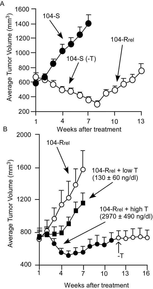
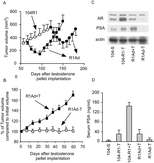
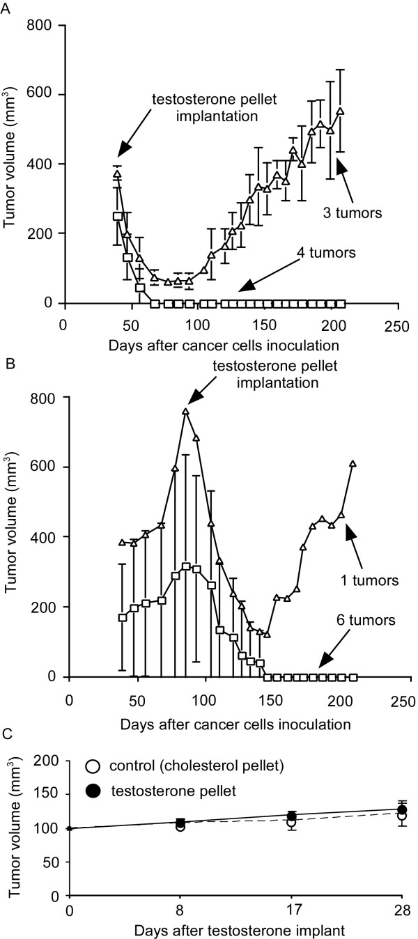
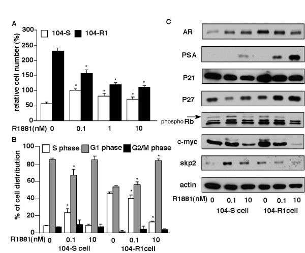
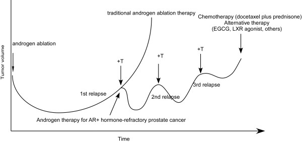
Prostate cancer is the most frequently diagnosed non-cutaneous tumor of men in Western countries. While surgery is often successful for organ-confined prostate cancer, androgen ablation therapy is the primary treatment for metastatic prostate cancer. However, this therapy is associated with several undesired side-effects, including increased risk of cardiovascular diseases. Shortening the period of androgen ablation therapy may benefit prostate cancer patients. Intermittent Androgen Deprivation therapy improves quality of life, reduces toxicity and medical costs, and delays disease progression in some patients. Cell culture and xenograft studies using androgen receptor (AR)-positive castration-resistant human prostate cancers cells (LNCaP, ARCaP, and PC-3 cells over-expressing AR) suggest that androgens may suppress the growth of AR-rich prostate cancer cells. Androgens cause growth inhibition and G1 cell cycle arrest in these cells by regulating c-Myc, Skp2, and p27Kip via AR. Higher dosages of testosterone cause greater growth inhibition of relapsed tumors. Manipulating androgen/AR signaling may therefore be a potential therapy for AR-positive advanced prostate cancer.


Figures

Figure 1
Figure 1
Progression of hormone-dependent LNCaP 104-S tumors to androgen-ablation-resistant 104-Rrel tumors, and androgenic growth suppression of 104-Rrel tumors. (A) Mice were injected subcutaneously with hormone-dependent 104-S cells. After allowing tumors to grow for 7 weeks, mice were separated into control (filled circles, 14 mice with 19 tumors) and castration groups (open circles, 24 mice with 36 tumors) and the time was designated as week 1 [18]. (B) Mice in the castrated group in (A) at the 14th week were separated into 3 groups including a control group (open circles, 6 mice with 9 tumors), a low dosage testosterone treatment group that received a subcutaneous implant of a 20 mg Testosterone/cholesterol (1:9) pellet (filled squares, 9 mice with 12 tumors), and a high-dosage testosterone treatment group that received a subcutaneous implant of a 20 mg pure Testosterone pellet (filled circles, 10 mice with 12 tumors) [18]. Tumor volumes are expressed as the mean + standard error.
Figure 2
Figure 2
Progression and regression of LNCaP 104-R1 tumor xenografts in nude mice treated with testosterone. (A) LNCaP 104-R1 tumor xenografts in castrated male nude mice were allowed to grow until they reached an average volume of 300 mm3 on the 58th day. On the 67th day, mice were separated into a control group (open circles) and a treatment group (filled circles). The treatment group received a subcutaneous implant of a 20 mg testosterone pellet. The mice in the control group were implanted with a 20 mg testosterone pellet on day 121. Open circles represent tumor in mice without testosterone, while filled circles and filled squares represent tumors in mice with testosterone. Tumor volumes are expressed as the mean ± standard error [15]. (B) For mice carrying adapted R1Ad tumors from (A), testosterone pellets were removed from 5 mice (10 tumors). Their tumor growth was compared with tumors in mice bearing testosterone pellets (5 mice with 10 tumors) [15]. (C) PSA, AR, and actin protein levels in 104-S tumor (in intact mice), 104-R1-T tumors, R1Ad-1+T tumors, and R1Ad-T were assayed by Western blot [15]. (D) Serum PSA level of mice with 104-S tumors (in intact mice), 104-R1-T tumors, 104-R1+T tumors, R1Ad+T tumors, R1Ad-T tumors was determined by ELISA [15].
Figure 3
Figure 3
Regression and relapse of LNCaP CDXR-3 tumor xenografts in nude mice treated with testosterone LNCaP CDXR tumor xenografts in castrated male nude mice were allowed to grow until they reached an average volume of 400 mm3 on the 38th day. All mice carrying tumors received a subcutaneous implant of a 20 mg testosterone pellet. The mice in the control group were implanted with a 20 mg testosterone pellet either at an early stage (50 days after inoculation, 7 tumors) (A) or late stage (92 days after inoculation, 7 tumors) (B) [27]. Open triangles represent tumors relapsed, while open squares represent tumors disappeared after androgen treatment. Tumor volumes are expressed as the mean ± standard error. (C) LNCaP IS-3 xenogarfts were separated into control group (20 mg cholesterol pellet implant, 9 tumors) and treatment group (20 mg testosterone pellet implant, 10 tumors) to determine the effect of androgen on growth of IS tumors [21].
Figure 4
Figure 4
Effect of androgen on cell proliferation, cell cycle, and cell cycle-related proteins in hormone-dependent 104-S and androgen ablation-resistant 104-R1 cells. (A) LNCaP 104-S and 104-R2 cells were treated with increasing concentration of R1881 for 96 hours. Relative cell number was determined by using a 96-well proliferation assay and data were normalized to number of 104-S cells at 0.1 nM R1881. Asterisk (*) represents statistically significant difference between treatment group compared to control group of 104-S or 104-R1 cells. (B) Percentage of 104-S and 104-R1 cells in S phase determined by flow cytometry. LNCaP 104-S and 104-R2 cells were treated with increasing concentrations of R1881 for 96 hours. Values represent the mean + standard error derived from 5 independent experiments. (C) Protein expression of androgen receptor (AR), prostate specific antigen (PSA), p21cip, p27Kip, phosphor-retinoblastoma protein (Rb), c-Myc, S phase kinase-associated protein 2 (Skp2) were determined by Western blotting assay in 104-S and 104-R1 cells treated 96 hrs with different concentration of R1881. β-actin was used as loading control.
Figure 5
Figure 5
Androgen and alternative therapy for advanced prostate cancer. After androgen ablation therapy, androgen treatment will retard the growth and progression of AR-rich advanced tumors in patients. In that case, chemotherapy (docetaxel plus prednisone) or alternative therapies, such as EGCG, LXR agonist or other treatments, should be considered to suppress tumor growth.

References

    1. Huggins C, Stevens R, Hodges C. Studies on prostatic cancer: II. The effects of castration on advanced carcinoma of the prostate gland. Arch Surg. 1941;43:15.
    1. Sadar MD. Small molecule inhibitors targeting the "achilles' heel" of androgen receptor activity. Cancer Res. 2011;71:1208–1213. doi: 10.1158/0008-5472.CAN_10-3398. - DOI - PMC - PubMed
    1. Ibrahim T, Flamini E, Mercatali L, Sacanna E, Serra P, Amadori D. Pathogenesis of osteoblastic bone metastases from prostate cancer. Cancer. 2010;116:1406–1418. doi: 10.1002/cncr.24896. - DOI - PubMed
    1. Keller ET, Zhang J, Cooper CR, Smith PC, McCauley LK, Pienta KJ, Taichman RS. Prostate carcinoma skeletal metastases: cross-talk between tumor and bone. Cancer Metastasis Rev. 2001;20:333–349. doi: 10.1023/A:1015599831232. - DOI - PubMed
    1. Bubendorf L, Schopfer A, Wagner U, Sauter G, Moch H, Willi N, Gasser TC, Mihatsch MJ. Metastatic patterns of prostate cancer: an autopsy study of 1,589 patients. Hum Pathol. 2000;31:578–583. doi: 10.1053/hp.2000.6698. - DOI - PubMed

Publication types

MeSH terms

Substances