. 2012 Jan;71(1):92-8.
doi: 10.1136/ard.2011.151712. Epub 2011 Aug 21.

Peptidylarginine deiminase 2, 3 and 4 have distinct specificities against cellular substrates: novel insights into autoantigen selection in rheumatoid arthritis

Affiliations

Peptidylarginine deiminase 2, 3 and 4 have distinct specificities against cellular substrates: novel insights into autoantigen selection in rheumatoid arthritis

Erika Darrah et al. Ann Rheum Dis. 2012 Jan.

Abstract

Objective: To define the relationship between autoantigen citrullination and different peptidylarginine deiminase (PAD) enzymes in rheumatoid arthritis (RA).

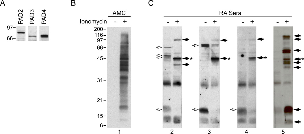
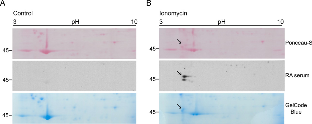
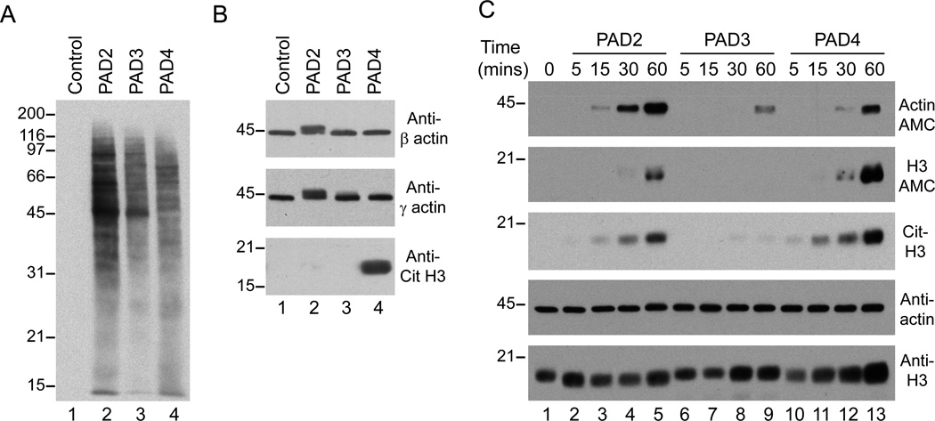
Methods: Citrullinated autoantigens were identified by immunoblotting control and ionomycin-activated human primary neutrophil lysate with RA sera. Autoantigen identity and citrullination sites were defined by mass spectrometry. PAD isoenzyme expression in human neutrophils was determined by immunoblotting. PAD substrate specificity was addressed in HL-60 cell lysates co-incubated with human recombinant PAD2, PAD3 and PAD4.

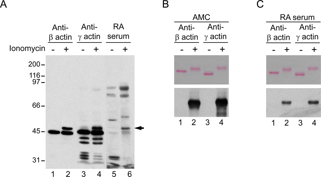
Results: Although prominent protein citrullination is observed in ionomycin-activated neutrophils, RA sera only recognised a limited number of these citrullinated molecules. Among these, the authors identified that β and γ-actins are citrullinated on at least 10 arginine residues, generating a novel 47 kDa species that is frequently recognised by RA autoantibodies. Interestingly, the authors showed that the PAD enzymes expressed in human neutrophils (ie, PAD2, PAD3 and PAD4) have unique substrate specificities, independent of their subcellular distribution. Thus, only PAD2 was able to citrullinate native β/γ-actin, while histone H3 was only citrullinated by PAD4.

Conclusion: These studies identified β and γ-actins as novel citrullinated autoantigens in RA, allowing enzyme specificity against intracellular substrates to be addressed. The studies provide evidence that PAD enzymes have the intrinsic capacity to select unique protein targets. The authors propose that unique PAD specificity may play a role in autoantigen selection in RA.


Figures

Figure 1
Figure 1
PAD expression in human primary neutrophils and autoantigen recognition by RA sera in control and ionomycin-activated neutrophils. A. PAD2, 3 and 4 are expressed in human neutrophils. Samples from freshly isolated neutrophils were analyzed by immunoblotting with antibodies against human PAD2, PAD3 and PAD4. B, C. Primary human neutrophils in HBSS containing 2 mM CaCl2 were incubated in the absence (−) or presence (+) of 1 µM ionomycin for 4 hrs at 37°C. Samples were analyzed by electrophoresis on 13% SDS-polyacrylamide gels and immunoblotted using an AMC antibody (B) or anti-CCP positive sera from RA patients (C). Data from 4 representative sera are shown in C. The unfilled arrows denote antigens detected in non-stimulated neutrophils, filled arrows mark antigens generated upon neutrophil activation and the asterisk denotes the 47kDa species that was further analyzed by mass spectrometry.
Figure 2
Figure 2
Two-dimentional mapping of the 47kDa RA autoantigen generated in ionomycin-activated neutrophils. Primary human neutrophils in HBSS containing 2 mM CaCl2 were incubated in the absence (A) or presence (B) of 1 µM ionomycin for 4 hrs at 37°C. Samples were resolved in the first dimension using 3–10 IPG strips and then by electrophoresis on 8% SDS-polyacrylamide gels. Proteins were visualized by Ponceau S staining (upper panel) prior to immunoblotting with RA patient sera (middle panel). Data from one representative serum is shown. Once the antigen was mapped, the spot was sliced from a GelCode blue-stained gel (lower panel) and used for protein sequencing. Note that the 47kDa species detected by RA sera (marked with the black arrow) is absent in control cells.
Figure 3
Figure 3
Beta and gamma actin are citrullinated autoantigens in RA. A. Beta- and gamma-actin are modified in ionomycin activated neutrophils and the novel species co-migrates with the 47kDa RA autoantigen. Samples from control (lanes 1, 3, and 5) and ionomycin activated neutrophils (lanes 2, 4 and 6) were analyzed by immunoblotting using monoclonal antibodies against human beta- and gamma-actin (Sigma) or RA serum. The filled arrow denotes the 47kDa species detected by anti-actin antibodies and RA sera. B, C. Beta- and gamma-actin are citrullinated and recognized by RA autoantibodies. Recombinant human beta-actin (lanes 1 and 2) or gamma-actin (lanes 3 and 4) was incubated in the absence (lanes 1 and 3) or presence (lanes 2 and 4) of rabbit PAD2. After 1 hr at 37°C, samples were divided in two and analyzed by electrophoresis on 8% SDS-polyacrylamide gels. After electrophoresis, proteins were visualized by Ponceau S staining (upper panels) prior to AMC immunoblotting (B, lower panel) or immunoblotting with RA sera (C, lower panel). Data from 1 representative serum containing anti-citrullinated actin antibodies is shown in C.
Figure 4
Figure 4
PAD2, 3 and 4 have distinct substrate specificities. A, B. Sonicated HL-60 cell lysates were incubated in the absence (lane 1) or presence of human rPAD2, rPAD3 or rPAD4 (lanes 2–4, respectively) for 1 hr at 37°C. After terminating the reactions, samples were analyzed by electrophoresis on 13% SDS-polyacrylamide gels and immunoblotted with AMC antibodies (A) or antibodies against human beta-actin, gamma-actin (Sigma) and citrullinated histone H3 (citruline 2 + 8 + 17) (Abcam) (B). C. A mixture of purified human actin plus recombinant histone H3 was incubated in the absence (lane 1) or presence of human rPAD2 (lanes 2–5), rPAD3 (lanes 6–9) or rPAD4 (lanes 10–13) for 5–60 mins at 37°C. After terminating the reactions, samples were analyzed in duplicated (actin) or triplicated (histone H3) by electrophoresis on 13% SDS-polyacrylamide gels. After electrophoresis, proteins were visualized by immunoblotting with antibodies against AMC, citrullinated histone H3 (citrulline 2 + 8 + 17) (Cit-H3), human beta/gamma-actin and histone H3 (Abcam).
Figure 5
Figure 5
Cirullination sites in actin. A, B and C. Amino acid sequence (gamma-actin in A and beta-actin in B) and tertiary structure (C) of actin. The structure of actin consists of two domains called the large and small domains (shown in blue and orange, respectively), with a cleft containing the bound nucleotide (marked as ATP in C). The amino acid sequence covered by mass spectrometry analysis of endogenous (A) and recombinant (B) citrullinated actin is shown in red, the arginines that are not citrullinated are shown in green and the potential arginine targets for citrullination are shown in yellow. The tertiary structure of actin (C) reveals clustering of citrullination sites around the cleft and the surface of the large domain opposite to the cleft region. The model shown in C was generated from the Molecular Modeling DataBase (NCBI) using the software Cn3D according to coordinates generated by Schutt et al.

Similar articles

Cited by

References

    1. Vossenaar ER, Zendman AJ, van Venrooij WJ, et al. Pad, a growing family of citrullinating enzymes: genes, features and involvement in disease. BioEssays. 2003;25:1106–1118. - PubMed
    1. Wegner N, Lundberg K, Kinloch A, et al. Autoimmunity to specific citrullinated proteins gives the first clues to the etiology of rheumatoid arthritis. Immunol Rev. 2010;233:34–54. - PubMed
    1. Foulquier C, Sebbag M, Clavel C, et al. Peptidyl arginine deiminase type 2 (PAD-2) and PAD-4 but not PAD-1, PAD-3, and PAD-6 are expressed in rheumatoid arthritis synovium in close association with tissue inflammation. Arthritis Rheum. 2007;56:3541–3553. - PubMed
    1. Chang X, Yamada R, Suzuki A, et al. Localization of peptidylarginine deiminase 4 (PADI4) and citrullinated protein in synovial tissue of rheumatoid arthritis. Rheumatology (Oxford) 2005;44:40–50. - PubMed
    1. Kinloch A, Lundberg K, Wait R, et al. Synovial fluid is a site of citrullination of autoantigens in inflammatory arthritis. Arthritis Rheum. 2008;58:2287–2295. - PubMed

Publication types

MeSH terms