. 2011 Nov;32(11):1689-96.
doi: 10.1093/carcin/bgr191. Epub 2011 Aug 22.

Inhibition of chronic pancreatitis and pancreatic intraepithelial neoplasia (PanIN) by capsaicin in LSL-KrasG12D/Pdx1-Cre mice

Affiliations

Inhibition of chronic pancreatitis and pancreatic intraepithelial neoplasia (PanIN) by capsaicin in LSL-KrasG12D/Pdx1-Cre mice

Han Bai et al. Carcinogenesis. 2011 Nov.

Abstract

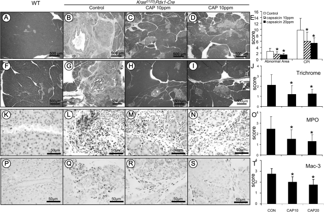
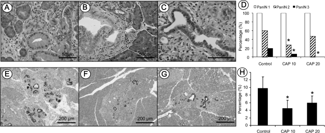
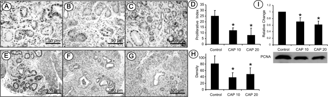
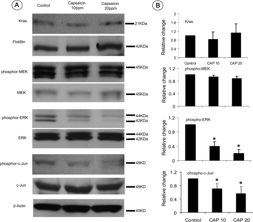
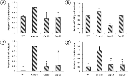
Capsaicin is a major biologically active ingredient of chili peppers. Extensive studies indicate that capsaicin is a cancer-suppressing agent via blocking the activities of several signal transduction pathways including nuclear factor-kappaB, activator protein-1 and signal transducer and activator of transcription 3. However, there is little study on the effect of capsaicin on pancreatic carcinogenesis. In the present study, the effect of capsaicin on pancreatitis and pancreatic intraepithelial neoplasia (PanIN) was determined in a mutant Kras-driven and caerulein-induced pancreatitis-associated carcinogenesis in LSL-Kras(G12D)/Pdx1-Cre mice. Forty-five LSL-Kras(G12D)/Pdx1-Cre mice and 10 wild-type mice were subjected to one dose of caerulein (250 μg/kg body wt, intraperitoneally) at age 4 weeks to induce and synchronize the development of chronic pancreatitis and PanIN lesions. One week after caerulein induction, animals were randomly distributed into three groups and fed with either AIN-76A diet, AIN-76A diet containing 10 p.p.m. capsaicin or 20 p.p.m. capsaicin for a total of 8 weeks. The results showed that capsaicin significantly reduced the severity of chronic pancreatitis, as determined by evaluating the loss of acini, inflammatory cell infiltration and stromal fibrosis. PanIN formation was frequently observed in the LSL-Kras(G12D)/Pdx1-Cre mice. The progression of PanIN-1 to high-grade PanIN-2 and -3 were significantly inhibited by capsaicin. Further immunochemical studies revealed that treatment with 10 and 20 p.p.m. capsaicin significantly reduced proliferating cell nuclear antigen-labeled cell proliferation and suppressed phosphorylation of extracellular signal-regulated kinase (ERK) and c-Jun as well blocked Hedgehog/GLI pathway activation. These results indicate that capsaicin could be a promising agent for the chemoprevention of pancreatic carcinogenesis, possibly via inhibiting pancreatitis and mutant Kras-led ERK activation.


Figures

Fig. 1.
Fig. 1.
Analysis of chronic pancreatitis using the approaches of histopathology, immunohistochemistry and histochemistry. Histopathology in the pancreatic tissue of KrasG12D/Pdx1-Cre mice: (A) Pancreas in wild-type control mice showing morphologically normal pancreatic parenchyma (acini and islets as well interlobular ducts). (B) Extensive chronic pancreatitis in KrasG12D/Pdx1-Cre mice fed AIN-76A diet alone showing loss of acini and stromal fibrosis. (C and D) Minimal to mild chronic pancreatitis in mice fed 10 or 20 p.p.m. capsaicin-supplemented diet, respectively. (E) Semiquantitative analysis (histogram) of chronic pancreatitis based on the extent of acinar loss, stromal fibrosis and inflammatory cell infiltration. There was a statistically significant difference in mice fed AIN6A diet alone compared with those given capsaicin-supplemented diets (P < 0.05). Trichrome stain highlighting stromal fibrosis in the pancreas: (F) Trichrome stained fibroconnective tissue (blue color) only in the interlobular areas of a normal pancreas in wild-type control mice. (G) KrasG12D/Pdx1-Cre mice fed AIN-76A diet alone showing extensive fibrosis in pancreatic parenchyma. (H and I) mice fed 10 or 20 p.p.m. capsaicin-supplemented diet exhibiting much less stromal fibrosis in the pancreas. (J) Semiquantitative analysis (histogram) of the extent of trichrome stain-labeled stromal fibrosis revealing statistically significant differences in mice fed capsaicin-supplemented diets compared with those given AIN-76A diet alone (P < 0.05). MPO-labeled neutrophils in the pancreas: (K) Pancreas from wild-type control mice showing no MPO-labeled neutrophils. (L) KrasG12D/Pdx1-Cre mice fed AIN-76A diet alone displaying intense MPO-labeled neutrophils in the areas of pancreatitis. (M and N) Mice fed 10 or 20 p.p.m. capsaicin-supplemented diet exhibiting decreased MPO-positive neutrophils. (O) Semiquantitative analysis (histogram) of the MPO-labeled inflammatory cells showing a statistically significant difference in mice fed capsaicin-supplemented diets compared with those given AIN-76A diet alone (P < 0.05). Mac-3-labeled macrophages in the pancreas: (P) Pancreas from wild-type control mice showing no Mac-3-labeled inflammatory cells. (Q) KrasG12D/Pdx1-Cre mice fed AIN-76A diet alone displaying intense Mac-3-labeled macrophage in the areas of pancreatitis. (R andS) Mice fed 10 or 20 p.p.m. capsaicin-supplemented diet exhibiting decreased Mac-3-positive macrophages. (T) Semiquantitative analysis (histogram) of the Mac-3-labeled macrophages showing a statistically significant difference in mice fed capsaicin-supplemented diets compared with those given AIN-76A diet alone (P < 0.05).
Fig. 2.
Fig. 2.
Histopathological and histochemical analysis of mPanIN lesions in KrasG12D/Pdx1-Cre mice. (AC) Representative photos of mPanIN-1, mPanIN-2 and mPanIN-3, respectively. (D) Semiquantitative analysis (histogram) of mPanIN lesions. Statistically significant lower in the incidence of high-grade mPanIN-2 and mPanIN-3 lesions were observed in mice fed capsaicin-supplemented diets compared with AIN-76A diet alone (Chi-square test, P < 0.05). Alcian blue/PAS staining of mucin in mPanIN lesions in (E) mice fed AIN-76A diet alone, or (F and G) mice fed capsaicin-supplemented diets. (H) Statistical analysis of PAS/Alcian blue-positive staining areas in the control and capsaicin treatment groups (Chi-square test, P < 0.05).
Fig. 3.
Fig. 3.
Immunohistochemistry of PCNA-labeled cell proliferation and phosopho-ERK expression. (A) PCNA-labeled cell proliferation in KrasG12D/Pdx1-Cre mice fed AIN-76A diet alone. (B and C) Mice fed capsaicin-supplemented diets. (D) Semiquantitative analysis (histogram) of PCNA-labeled cell proliferation. Statistical significance was observed in mice fed capsaicin-supplemented diets compared with those given AIN-76A diet alone (P < 0.05). (E) Phospho-ERK in KrasG12D/Pdx1-Cre mice fed AIN-76A diet alone. (F and G) Phospho-ERK in mice fed capsaicin-supplemented diets. (H) Semiquantitative analysis (histogram) of phospho-ERK staining intensity revealed statistically significant differences in mice fed capsaicin-supplemented diets compared with those given AIN-76A diet alone (P < 0.05). (I) Western blot of PCNA showing the protein level is statistically lower in the pancreas of KrasG12D/Pdx1-Cre mice fed capsaicin-supplemented diets compared with those fed AIN6A diet alone (P < 0.05).
Fig. 4.
Fig. 4.
Western blot assay of mutant Kras and its activated downstream signals phospho-MEK, phospho-ERK and Phospho-c-Jun. (A) Expression levels of Kras and its downstream phosphorylated and non-phosphorylated MEK, ERK and c-Jun in the pancreatic tissues detected using a western blot assay. (B) Histograms reveal the intensity of expression of membrane-bound Kras and each phospho-protein in mice fed AIN-76A diet alone or capsaicin-supplemented diets (*P < 0.05).
Fig. 5.
Fig. 5.
Quantitative PCR analysis of mRNA expression of TGF-β, PDGF-β, Shh and GLI1 in the pancreatic tissue in wild-type control mice (WT) and LSL-KrasG12D/Pdx1-Cre mice fed a diet with or without capsaicin. (A) TGF-β; (B) PDGF-β; (C) Shh and (D) GLI1 (*, Significant difference between mice with or without capsaicin treatment, P < 0.05).

References

    1. Whitcomb DC. Inflammation and Cancer V. Chronic pancreatitis and pancreatic cancer. Am. J. Physiol. Gastrointest. Liver. Physiol. 2004;287:G315–G319. - PubMed
    1. Hussain SP, et al. Radical causes of cancer. Nat. Rev. Cancer. 2003;3:276–285. - PubMed
    1. Hermanova M, et al. Expression of COX-2 is associated with accumulation of p53 in pancreatic cancer: analysis of COX-2 and p53 expression in premalignant and malignant ductal pancreatic lesions. Eur. J. Gastroenterol. Hepatol. 2008;20:732–739. - PubMed
    1. Whitcomb D, et al. Germ-line mutations, pancreatic inflammation, and pancreatic cancer. Clin. Gastroenterol. Hepatol. 2009;7:S29–S34. - PubMed
    1. Papoiu AD, et al. Topical capsaicin. The fire of a ‘hot' medicine is reignited. Expert Opin. Pharmacother. 2010;11:1359–1371. - PubMed

Publication types

MeSH terms