. 2011 Aug 22;194(4):597-612.
doi: 10.1083/jcb.201102018.

Ran-dependent docking of importin-beta to RanBP2/Nup358 filaments is essential for protein import and cell viability

Affiliations

Ran-dependent docking of importin-beta to RanBP2/Nup358 filaments is essential for protein import and cell viability

Masakazu Hamada et al. J Cell Biol. .

Abstract

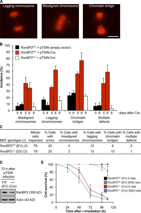
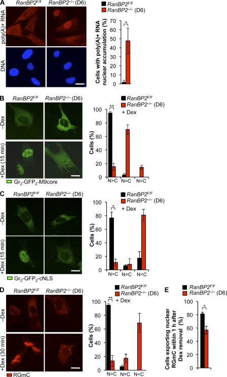
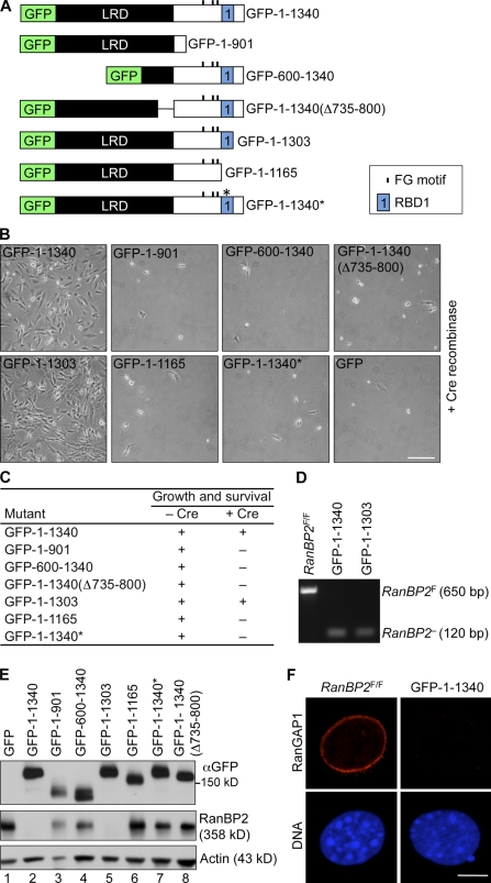
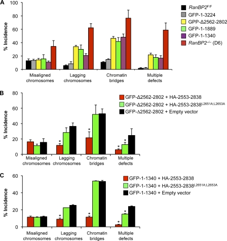
RanBP2/Nup358, the major component of the cytoplasmic filaments of the nuclear pore complex (NPC), is essential for mouse embryogenesis and is implicated in both macromolecular transport and mitosis, but its specific molecular functions are unknown. Using RanBP2 conditional knockout mouse embryonic fibroblasts and a series of mutant constructs, we show that transport, rather than mitotic, functions of RanBP2 are required for cell viability. Cre-mediated RanBP2 inactivation caused cell death with defects in M9- and classical nuclear localization signal (cNLS)-mediated protein import, nuclear export signal-mediated protein export, and messenger ribonucleic acid export but no apparent mitotic failure. A short N-terminal RanBP2 fragment harboring the NPC-binding domain, three phenylalanine-glycine motifs, and one Ran-binding domain (RBD) corrected all transport defects and restored viability. Mutation of the RBD within this fragment caused lethality and perturbed binding to Ran guanosine triphosphate (GTP)-importin-β, accumulation of importin-β at nuclear pores, and cNLS-mediated protein import. These data suggest that a critical function of RanBP2 is to capture recycling RanGTP-importin-β complexes at cytoplasmic fibrils to allow for adequate cNLS-mediated cargo import.


Figures

Figure 1.
Figure 1.
Generation of MEFs lacking RanBP2. (A) An overview of the knockout strategy. Relevant portions of the RanBP2 hypomorphic allele (H), the floxed allele (F), and the null allele (−) are shown. (B) Images of RanBP2F/F MEF cultures taken 8 d after infection with empty or Cre-containing lentivirus. (C) Western blots of MEF extracts prepared at the indicated times after infection with Cre lentivirus. (D) Images illustrating that NPCs of RanBP2−/− (D6) MEFs lack RanBP2. (E) Images showing that RanGAP1 is unable to localize to nuclear pores in RanBP2−/− (D6) MEFs. (F) Images showing that SUMO1 fails to decorate the nuclear envelope in RanBP2−/− (D6) MEFs. (G) Western blots of MEF extracts of RanBP2F/F and RanBP2−/− (D6) MEFs probed for RanGAP1. Actin served as a loading control. Bars: (B) 500 µm; (D–F) 5 µm.
Figure 2.
Figure 2.
High chromosome missegregation rates but no mitotic catastrophe in MEFs lacking RanBP2. (A) Representative chromosome segregation errors in RanBP2−/− (D6) MEFs. Images are from live H2B-mRFP–expressing cells. Bar, 10 µm. (B) Analysis of chromosome segregation errors at days 5 and 6 after infecting RanBP2F/F MEFs with empty or Cre-containing lentivirus. RanBP2+/+ MEFs were used to control for chromosome missegregation that might result from expression of Cre recombinase (Loonstra et al., 2001). Error bars represent SEM. Three independent lines were analyzed per genotype/per treatment (z25 cells per line, z75 total per genotype/per treatment). (C) Chromosome segregation errors at 72 h after infecting RanBP2F/F MEFs with empty or Cre-containing lentivirus. EV, empty vector. (D) Western blot analysis of MEF extracts of RanBP2F/F and RanBP2−/− (D3) MEFs probed for RanBP2. Actin served as a loading control. (E) Survival curves of RanBP2-null and control MEFs with and without γ irradiation. RanBP2F/F MEFs were infected with empty or Cre-containing lentivirus and selected with puromycin for 24 h. Cells were then seeded in dishes and either not irradiated or exposed to 3,000 rads of γ irradiation at 72 h after infection (t = 0 in the graph). Cells were counted at 24-h intervals. n = 5 lines per genotype. Error bars represent SD.
Figure 3.
Figure 3.
Multiple transport pathways are perturbed in RanBP2-null MEFs. (A) Measurement of poly(A)+ RNA export from the nucleus (n = 3 MEF lines per genotype; >50 cells per line). (B) Measurement of protein import using the Gr2-GFP2-M9 reporter. Three independently generated cell lines were used (RanBP2F/F, n = 168; RanBP2−/− [D6], n = 97). (C) Measurement of protein import using the Gr2-GFP2-cNLS reporter. Three independently generated cell lines were used (RanBP2F/F, n = 167; RanBP2−/− [D6], n = 157). (D and E) Combined measurement of NLS protein import (D) and NES export (E) using RGmC. Three independently generated cell lines were used. (D) RanBP2F/F, n = 188; RanBP2−/− [D6], n = 173. (E) RanBP2F/F, n = 305; RanBP2−/− [D6], n = 332. (A–D) *, P < 0.01; **, P < 0.001 (two-tailed unpaired t test). Error bars indicate SD. C, cytoplasm; N, nucleus. Bars, 10 µm.
Figure 4.
Figure 4.
The RanBP2 SUMO E3 ligase domain is not essential for cell viability. (A) An overview of wild-type and mutant RanBP2 proteins. The LRD includes the NPC-targeting domain. CY, cyclophilin homology domain; E3, SUMO E3 ligase domain; KBD, kinesin-binding domain; ZnFs, zinc finger motifs. (B) Fluorescent images of RanBP2−/− (D6) MEFs containing the indicated GFP-tagged RanBP2 mutants. Bar, 5 µm. (C) Images of RanBP2−/− (D8) MEFs containing the indicated expression constructs. Bar, 200 µm. (D) Viability of RanBP2F/F MEFs expressing the indicated RanBP2 proteins in the presence or absence of endogenous RanBP2. (E) PCR genotyping of RanBP2−/− MEFs expressing the indicated RanBP2 proteins showing complete absence of RanBP2F alleles. (F) Western blot analysis of MEFs carrying the indicated GFP-tagged RanBP2 expression constructs. Actin was used as a loading control. MEFs in the first three lanes were Cre treated and lacked endogenous RanBP2, whereas MEFs in the last two lanes were not treated with Cre (nonsurviving mutants). The antibody against RanBP2 did not detect GFP-1–1889 and GFP-Δ2562–2802 proteins because these mutants lack the antibody epitope (Dawlaty et al., 2008). Asterisks mark endogenous RanBP2 protein.
Figure 5.
Figure 5.
The N terminus of RanBP2 is sufficient for cell growth and survival. (A) An overview of mutant RanBP2 proteins. 1–1340*, 1–1340 containing a W1211R and K1212M double mutation in RBD1 designed to disrupt Ran binding and potentiation of RanGAP1. (B) Images of RanBP2−/− (D8) MEFs containing the indicated expression constructs. Bar, 200 µm. (C) Viability of RanBP2F/F MEFs expressing the indicated RanBP2 proteins in the presence or absence of endogenous RanBP2. (D) PCR analysis of DNA isolated from viable RanBP2−/− (D6) MEFs expressing the indicated RanBP2 proteins. (E) A Western blot of MEFs carrying the indicated GFP-tagged RanBP2 expression constructs. Actin was used as a loading control. MEFs in lanes 2 and 5 were Cre treated (lacking endogenous RanBP2), whereas MEFs in the remaining lanes were untreated. (F) RanBP2−/− MEFs expressing GFP-RanBP2-1–1340 and control MEFs stained for RanGAP1. Bar, 5 µm.
Figure 6.
Figure 6.
The RanBP2 SUMO E3 ligase domain acts autonomously in mitosis. (A) An analysis of segregation errors in RanBP2−/− (D6) MEFs and RanBP2−/− MEFs expressing the indicated RanBP2 mutants (n = 3 MEF lines per mutant; ∼25 cells per line). Error bars indicate SEM. All mutants are P < 0.05 versus RanBP2−/− (D6) MEFs (χ2 test). (B) An incidence of chromosome segregation errors in GFP-RanBP2-Δ2562–2802 RanBP2−/− MEFs expressing wild-type or mutant SUMO E3 ligase domain (n = 3 MEF lines evaluated for each mutant combination; ∼20–25 cells per line). (C) An incidence as described in B but for GFP-RanBP2-1–1340 RanBP2−/− MEFs. (B and C) *, P < 0.05 versus GFP-RanBP2-Δ2562–2802 RanBP2−/− MEFs carrying empty expression vector (χ2 test). Error bars indicate SEM.
Figure 7.
Figure 7.
cNLS-mediated import selectively requires Ran binding ability. (A) An analysis of poly(A)+ RNA distribution in RanBP2−/− (D6) MEFs expressing the indicated RanBP2 mutants (n = 3 MEF lines per mutant). 1–1340*, 1–1340 containing a W1211R and K1212M double mutation in RBD1 designed to disrupt Ran binding and potentiation of RanGAP1. (B) Measurement of protein import using the Gr2-GFP2-cNLS reporter. C, cytoplasm; N, nucleus. (C) Measurement of protein import using the NES-GFP2-cNLS reporter. (D) In vitro import of FITC-labeled cNLS-BSA (at 50 mM importin-β). (A–C) Error bars indicate SD. (B and C) *, P < 0.01; **, P < 0.001 (two-tailed unpaired t test). n = 3 MEF lines per mutant genotype; ∼50 cells per line. Bars,10 µm.
Figure 8.
Figure 8.
Importin-β accumulation at the NPC is dependent on RanBP2 RBD1. (A) Images of RanBP2−/− (D6) MEFs expressing the indicated RanBP2 mutants immunostained for importin-β. Nuclei were visualized with Hoechst. 1–1340*, 1–1340 containing a W1211R and K1212M double mutation in RBD1 designed to disrupt Ran binding and potentiation of RanGAP1. (B) In vitro import of FITC-labeled NLS-BSA into RanBP2−/− (D6) MEFs expressing the indicated RanBP2 mutants. Import assays were conducted at a twofold higher importin concentration as described in Fig. 7 D. (C) Blot overlay assay measuring binding of importin-β to various N-terminal RanBP2 fragments in the absence or presence of RanGTP. (D) Images of digitonin-permeabilized RanBP2−/− MEFs expressing the indicated RanBP2 mutants stained for Crm1. Nuclei were visualized with Hoechst staining. (E) Quantification of Crm1 signal at the nuclear rim. Mean values of 10 cells per line are presented. Error bars represent SEM. **, P < 0.001 versus RanBP2−/− cells expressing GFP-1–3224 (two-tailed unpaired t test). Bars, 10 µm.

Similar articles

Cited by

References

    1. Arnaoutov A., Dasso M. 2005. Ran-GTP regulates kinetochore attachment in somatic cells. Cell Cycle. 4:1161–1165 10.4161/cc.4.9.1979 - DOI - PubMed
    1. Arnaoutov A., Azuma Y., Ribbeck K., Joseph J., Boyarchuk Y., Karpova T., McNally J., Dasso M. 2005. Crm1 is a mitotic effector of Ran-GTP in somatic cells. Nat. Cell Biol. 7:626–632 10.1038/ncb1263 - DOI - PubMed
    1. Aslanukov A., Bhowmick R., Guruju M., Oswald J., Raz D., Bush R.A., Sieving P.A., Lu X., Bock C.B., Ferreira P.A. 2006. RanBP2 modulates Cox11 and hexokinase I activities and haploinsufficiency of RanBP2 causes deficits in glucose metabolism. PLoS Genet. 2:e177 10.1371/journal.pgen.0020177 - DOI - PMC - PubMed
    1. Balciunas D., Wangensteen K.J., Wilber A., Bell J., Geurts A., Sivasubbu S., Wang X., Hackett P.B., Largaespada D.A., McIvor R.S., Ekker S.C. 2006. Harnessing a high cargo-capacity transposon for genetic applications in vertebrates. PLoS Genet. 2:e169 10.1371/journal.pgen.0020169 - DOI - PMC - PubMed
    1. Bastos R., Lin A., Enarson M., Burke B. 1996. Targeting and function in mRNA export of nuclear pore complex protein Nup153. J. Cell Biol. 134:1141–1156 10.1083/jcb.134.5.1141 - DOI - PMC - PubMed

Publication types

MeSH terms

Substances