. 2011 Oct;157(2):599-607.
doi: 10.1104/pp.111.181057. Epub 2011 Aug 22.

Structural resolution of the complex between a fungal polygalacturonase and a plant polygalacturonase-inhibiting protein by small-angle X-ray scattering

Affiliations

Structural resolution of the complex between a fungal polygalacturonase and a plant polygalacturonase-inhibiting protein by small-angle X-ray scattering

Manuel Benedetti et al. Plant Physiol. 2011 Oct.

Abstract

We report here the low-resolution structure of the complex formed by the endo-polygalacturonase from Fusarium phyllophilum and one of the polygalacturonase-inhibiting protein from Phaseolus vulgaris after chemical cross-linking as determined by small-angle x-ray scattering analysis. The inhibitor engages its concave surface of the leucine-rich repeat domain with the enzyme. Both sides of the enzyme active site cleft interact with the inhibitor, accounting for the competitive mechanism of inhibition observed. The structure is in agreement with previous site-directed mutagenesis data and has been further validated with structure-guided mutations and subsequent assay of the inhibitory activity. The structure of the complex may help the design of inhibitors with improved or new recognition capabilities to be used for crop protection.


Figures
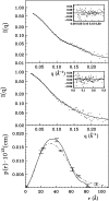
Figure 1.
Figure 1.
Experimental scattered intensities (dots) and the calculated ones with the ITP program relative to the complex between FpPG and PvPGIP2 (top section) and between FpPG and PvPGIP2.Q224K (middle section). The residuals are reported in the insets and the corresponding p(r) functions in the bottom section (continuous line for the complex between FpPG and PvPGIP2 and dot dash line for the complex between FpPG and PvPGIP2.Q224K). For clarity only a few error bars are shown.
Figure 2.
Figure 2.
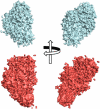
Consensus envelopes for the complex between FpPG and PvPGIP2 (cyan) and between FpPG and PvPGIP2.Q224K (red) as calculated with the GA_STRUCT program. Two orthogonal viewpoints are represented.
Figure 3.
Figure 3.
A, SDS-PAGE of the complex between FpPG and PvPGIP2 upon cross-linking with formaldehyde. A band of approximately 67 kD corresponding to the molecular mass of the complex accounted for more than 95% of the total proteins detectable on the gel. The cross-linking reaction was also performed in the presence of PvPGIP2 alone (approximately 31 kD; B) or FpPG alone (approximately 35 kD; C), to exclude the possibility that cross-linked homodimers or multimers are formed. In both cases, no high molecular weight bands were detected.
Figure 4.
Figure 4.
Experimental scattered intensities (dots) and the calculated ones with the ITP program relative to the complex between FpPG and PvPGIP2 in the presence of the cross-linker (top section). The residuals are reported in the insets and the corresponding p(r) function in the bottom section (dot dash line) in comparison with the p(r) function relative to the complex between FpPG and PvPGIP2 in the absence of the cross-linker (line). For clarity only a few error bars are shown.
Figure 5.
Figure 5.
Three-dimensional structure of the chemically cross-linked complex between FpPG (in green) and PvPGIP2 (in blue). A, The GA_STRUCT consensus envelope. B, The superimposition between the consensus envelope and the three-dimensional structure obtained with the SASREF code. C, The corresponding three-dimensional structures are shown. Two orthogonal viewpoints are reported.
Figure 6.
Figure 6.
A, Seven superimposed envelopes obtained for the chemically cross-linked complex between FpPG (green) and PvPGIP2 (blue) with the DAMAVER program. B, The superimposition between the consensus envelopes and the three-dimensional structures calculated with the SASREF code. C, The corresponding three-dimensional structures are shown. Two orthogonal viewpoints are reported.
Figure 7.
Figure 7.
Contact map of the residues involved in the FpPG-PvPGIP2 interaction. Contacts between residues of the two proteins are shown as squares in the map. The map was calculated using the SPACE server (http://ligin.weizmann.ac.il/cma/) and represents the average between the single contact maps obtained from the seven measurements and structures of Figure 6. The colors of the squares refer to the frequency with which each contact appears: yellow represents the presence of the contact in two out of seven structures; orange, red, green, blue, and black represent the presence of the contact in three, four, five, six, and seven out of seven structures, respectively. PvPGIP residues identified by Casasoli et al. (2009) as positively selected sites in Fabaceae and hotspots for the interaction with FpPG are indicated by a single and a double asterisk, respectively.

Similar articles

Cited by

References

    1. Bent AF, Mackey D. (2007) Elicitors, effectors, and R genes: the new paradigm and a lifetime supply of questions. Annu Rev Phytopathol 45: 399–436 - PubMed
    1. Bernadó P, Blanchard L, Timmins P, Marion D, Ruigrok RWH, Blackledge M. (2005) A structural model for unfolded proteins from residual dipolar couplings and small-angle x-ray scattering. Proc Natl Acad Sci USA 102: 17002–17007 - PMC - PubMed
    1. Boller T, Felix G. (2009) A renaissance of elicitors: perception of microbe-associated molecular patterns and danger signals by pattern-recognition receptors. Annu Rev Plant Biol 60: 379–406 - PubMed
    1. Bonivento D, Pontiggia D, Di Matteo A, Fernandez-Recio J, Salvi G, Tsernoglou D, Cervone F, Lorenzo GD, Federici L. (2008) Crystal structure of the endopolygalacturonase from the phytopathogenic fungus Colletotrichum lupini and its interaction with polygalacturonase-inhibiting proteins. Proteins 70: 294–299 - PubMed
    1. Brutus A, Sicilia F, Macone A, Cervone F, De Lorenzo G. (2010) A domain swap approach reveals a role of the plant wall-associated kinase 1 (WAK1) as a receptor of oligogalacturonides. Proc Natl Acad Sci USA 107: 9452–9457 - PMC - PubMed

Publication types

LinkOut - more resources