. 2012;195(5):377-91.
doi: 10.1159/000328712. Epub 2011 Aug 19.

Self-organization phenomena in embryonic stem cell-derived embryoid bodies: axis formation and breaking of symmetry during cardiomyogenesis

Affiliations

Self-organization phenomena in embryonic stem cell-derived embryoid bodies: axis formation and breaking of symmetry during cardiomyogenesis

Christiane Fuchs et al. Cells Tissues Organs. 2012.

Abstract

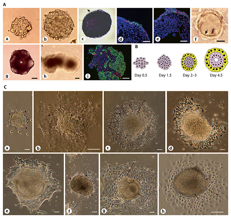
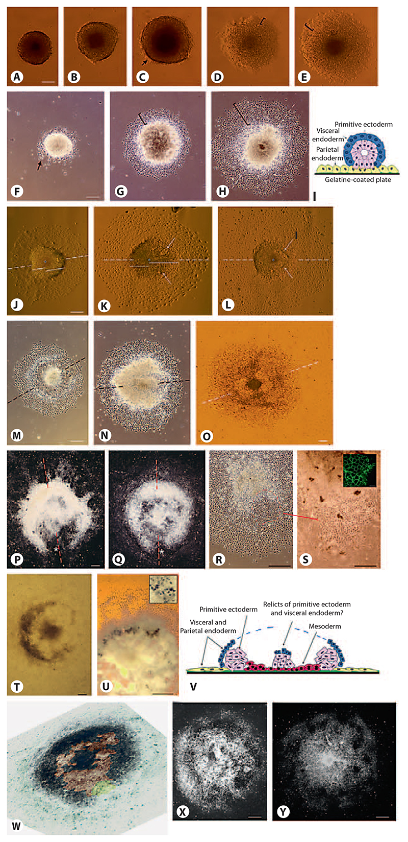
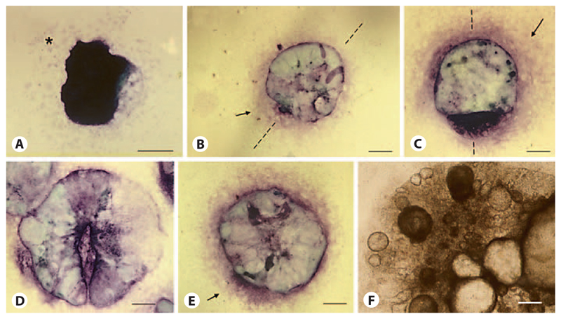
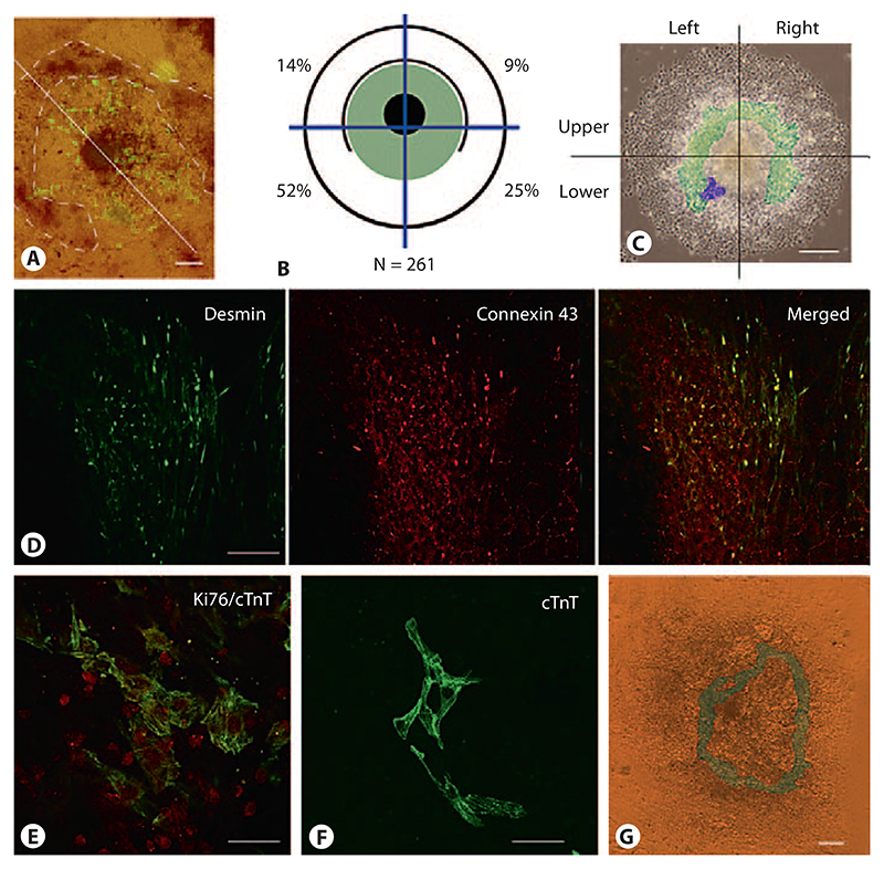
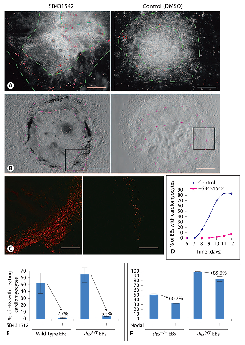
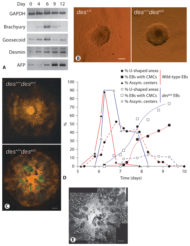
Aggregation of embryonic stem cells gives rise to embryoid bodies (EBs) which undergo developmental processes reminiscent of early eutherian embryonic development. Development of the three germ layers suggests that gastrulation takes place. In vivo, gastrulation is a highly ordered process but in EBs only few data support the hypothesis that self-organization of differentiating cells leads to morphology, reminiscent of the early gastrula. Here we demonstrate that a timely implantation-like process is a prerequisite for the breaking of the radial symmetry of suspended EBs. Attached to a surface, EBs develop a bilateral symmetry and presumptive mesodermal cells emerge between the center of the EBs and a horseshoe-shaped ridge of cells. The development of an epithelial sheet of cells on one side of the EBs allows us to define an 'anterior' and a 'posterior' end of the EBs. In the mesodermal area, first cardiomyocytes (CMCs) develop mainly next to this epithelial sheet of cells. Development of twice as many CMCs at the 'left' side of the EBs breaks the bilateral symmetry and suggests that cardiomyogenesis reflects a local or temporal asymmetry in EBs. The asymmetric appearance of CMCs but not the development of mesoderm can be disturbed by ectopic expression of the muscle-specific protein Desmin. Later, the bilateral morphology becomes blurred by an apparently chaotic differentiation of many cell types. The absence of comparable structures in aggregates of cardiovascular progenitor cells isolated from the heart demonstrates that the self-organization of cells during a gastrulation-like process is a unique feature of embryonic stem cells.


Figures

Fig. 1
Fig. 1. Radial symmetry of solitary EBs in hanging-drop cultures.
A 800 ESCs were aggregated in 20-μl drops for 4.5 days and then transferred to the suspension culture. ESCs aggregate within 1.5 days (a) and the compacted EB grows in size from day 2 to day 4.5 (b). At day 4.5 most cells are still AP-positive (c) with the exception of the outer layer of primitive endoderm (arrow). Primitive endoderm increasingly expresses Snail (red) and E-cadherin (green) (d, e). At the same time a Reichert’s membrane-like borderline (arrow) between the inner cells and the outer layer of primitive endoderm forms (f). If EBs were maintained in suspension culture the AP-positive cells became less from day 5 onwards (g) and a central cavity (*) formed, EBs developed mostly an irregular shape (h) and cells were distributed in an apparently chaotic manner as made evident by E-cadherin (green) and Snail (red) staining at day 10 (i). DNA was stained with DAPI (blue). a–i Bars: 100 μm. B Cartoon demonstrating the early development of cell types in EBs. Pink: ESCs and primitive ectodermal cells; yellow: primitive endoderm. C If EBs were transferred from the hanging drops to tissue culture plates at day 2 (a) or 3 (b) they did not further differentiate but remained as ESC-like colonies (arrowheads indicate area where primitive endoderm usually starts to grow and give rise to parietal endoderm; * = ESC-like cells). When plated on day 4.5, the primitive endoderm readily attached to the gelatine-coated tissue culture plate and differentiation proceeded with a radial symmetry (bracket in c). If plated on day 5.5, many EBs developed an irregular shape (d); however, differentiation proceeded normally in most cases 1–2 days later (e). If EBs were transferred to the tissue culture plate on day 6.5 or later, their development was either totally blocked (f, arrow-heads as in a) or only extraembryonic endoderm formed (brackets in g and h), while the inner cells remained entrapped within the Reichert’s membrane-like structure (arrows in g and h). Phase contrast images taken 1 day (a–d, f, h) and 2 days (e, g) after the attachment of EBs. Bars: 100 μm.
Fig. 2
Fig. 2. Attachment of EBs to a surface leads to the development of a bilateral symmetrical cell aggregate.
A–E Attachment of EBs to gelatine-coated tissue culture plates between days 4.5 and 6. C Arrow shows first attaching primitive endodermal cells. D, E Brackets show proliferating primitive endoderm. F, G Development of EBs between days 5 and 6. F Arrow shows migrating primitive endodermal cells. G, H Brackets show parietal endodermal cells undergoing an epithelial-mesenchymal transition. H In the meantime, the core of the EBs starts to elongate. I Cartoon: cross section of a typical EB at days 5 and 6. Inner cells of primitive ectoderm (pink) are surrounded by primitive endoderm (blue) which differentiates upon contact with the gelatine-coated tissue culture plate (green) into parietal endoderm (yellow) and visceral endoderm on top of the primitive ectoderm, respectively. J–L Breaking of the point symmetry by elongation of the central dense core of the EBs between day 6.5 and 7.5. Dashed lines indicate the forming axis. Solid lines emanating from the center (dots) of EBs indicate the asymmetric elongation of the EB core. K, L Arrows indicate areas where the thin cell layer (mesoderm?) forms which later separates the horse-shoe-shaped area from the center of the EBs. M–Q Development of the horseshoe-shaped ridge of cells surrounding the core of the EBs between days 7 and 8.5. R, S Epithelial sheet of cells developing at the open side of the horseshoe-shaped ridge at day 6.5. Dashed line demarcates epithelial sheet of cells; dot shows center of EB. Inset β-Catenin staining of epithelial cells (green); bar: 10 μ,m. T, U Hematopoietic cells develop inside of the horseshoe-shaped area of EBs. Inset Single erythrocyte-like cells; bar: 10 μ,m; benzidine stain. V Cartoon of EB at day 8 ± 1: transverse section through the center of the EB. W Pseudo-3-D image (tilted illumination under a stereomicroscope) of a day-8 EB where pseudocoloring indicates the area of developing presumptive mesoderm (brown), the epithelial sheet of cells at one end of the EB (yellow), and the dense horseshoe-shaped area of presumptive ectoderm (black) which is surrounded by the extraembryonic endoderm (green-blue). The central black area may be a remnant of the primitive endoderm covered by visceral endoderm. X, Y Blurring of the horseshoe-shaped ridge by cell growth and migration starting at day 8.5–9.0. Dashed lines indicate 2-fold axes. A–E, J–L, O, U, V Bright-field illumination. F–H, M, N, R, S Phase-contrast illumination. P, Q, X, Y Dark-field illumination. Bars: 100 μm except for S: 50 μm.
Fig. 3
Fig. 3
Development of the primitive endoderm after the attachment of EBs. Toluidine blue O staining of endodermal cells: epithelial primitive and visceral endoderm (dark purple), mesenchymal parietal endoderm (bright rose and arrows) [Bader et al., 2001]. Attached EB at day 5 (A), day 6 (B), day 7.5 (C), day 8.5 (D, E), day 12 (F). Bright-field image. Bars: 100 μm.
Fig. 4
Fig. 4. Development of first rhythmically contracting CMCs breaks the bilateral symmetry in EBs.
A Localization of CMCs between the horseshoe-shaped ridge of cells and the core of the EBs at day 9. Immunofluorescence staining of CMCs with MHC-alpha antibodies. Fluorescence image was overlaid onto the bright-field image. Dashed line demarcates the horseshoe-shaped ridge; the axis is indicated by a white line. B Schematic drawing of the EB composed of a dense center surrounded by a horseshoe-shaped dense ridge of cells. Distribution of the first rhythmically contracting CMCs in EBs is shown in the area highlighted in green, the black circle indicates the rim of the EB, blue lines demarcate the quadrants where the first contracting CMCs were located. Percentages of EBs wherein a single cluster of CMCs first started to contract are indicated. Total number of EBs monitored in 3 experiments was 373. C Area where first CMCs were located in EBs. False colors on top of a phase-contrast image of a typical EB at day 7.8: green shows the ‘gap-like’ area between the dense center of the EBs and the horseshoe-shaped ridge of cells, purple indicates the place where the majority of the first CMCs become visible. D Double-immunofluorescence staining of developing CMCs with Desmin (green) and Connexin 43 (red) antibodies. Note Connexin 43-positive and Desmin-negative epithelial cells at the lower right area of the image. E Double-immunofluores-cence staining of developing CMCs with Ki67 (red) and cardiac Troponin T (cTnT) antibodies (green). F Immunofluorescence staining of more mature CMCs with cTnT antibodies. G Ring-like network composed of rhythmically and synchronously contracting CMCs at day 12. Contracting cells are highlighted in green. Bright-field image. Bars: 100 μm (A, C, G), 20 μm (D, E), 40 μm (F).
Fig. 5
Fig. 5. Inhibition of Nodal-ALK4 signaling causes cell death in the area between the dense center and the horseshoe-shaped ridge in EBs.
A Day-7 EB cultured in the presence of 3.8 μmol/l SB421542 from day 3 to day 7. Dark-field image overlaid with red fluorescent image of propidium iodide-stained nuclei of dead cells. Dashed lines in A (green) and B (purple) demarcate area where mesoderm forms. B Day-11 EB cultured in the presence of 3.8 μmol/l SB421542 from day 3 to day 11. Note dark ring of dead cells in the presence of SB431542. Phase-contrast images. C Fluorescence image of propidium iodide staining of dead cells in areas indicated by rectangles in B. D Onset of cardiomyogenesis in EBs treated with SB431542. E Cardiomyogenesis in EBs in the presence of 3.8 μmol/l SB431542 in wild-type and desect EBs, respectively. Note increased percentage of EBs surviving SB431542 treatment in the presence of ectopically expressed Desmin. Same volume of DMSO was added to all controls as used to administer SB431542. F Cardiomyogenesis in des–/– and in desect EBs in the presence of 58 nmol/l recombinant Nodal. Data are from 2 experiments (error bars, standard deviation). p < 0.05. Number of EBs checked per experiment: 180. Bars: 200 μm (A), 100 μm (B), 50 μm (C).
Fig. 6
Fig. 6. Ectopic expression of the early muscle-specific protein Desmin disturbs morphogenesis in EBs.
A Semiquantitative RT-PCR analysis of mRNAs isolated from developing EBs at times indicated. GAPDH was loading control. Alpha fetoprotein (AFP) was used as control to monitor differentiation of endoderm. B Typical morphology of wild-type (des+/+) and des+/+ desect EBs at days 6–7. C des+/+desect EBs at days 8–9. D Development of asymmetric centers in EBs (▲), horseshoe-shaped ridges surrounding the centers (●), and rhythmically contracting CMCs (◼) in wild-type (red lines; n = 788) and des+/+ desect (blue lines; n = 706) EBs. Standard deviation was always less than 15% and is omitted for clarity reasons. E An aggregate of somatic stem cells isolated from murine hearts at day 13 after aggregation. B Phase-contrast images. C, E Dark-field pictures composed of several images. Areas with rhythmically contracting CMCs are highlighted in green. Bars: 200 μm (B), 100 μm (C), 1 mm (E).

Similar articles

Cited by

References

    1. Aubert J, Dessolin S, Belmonte N, Li M, McKenzie FR, Staccini L, Villageois P, Barhanin B, Vernallis A, Smith AG, Ailhaud G, et al. Leukemia inhibitory factor and its receptor promote adipocyte differentiation via the mitogen-activated protein kinase cascade. J Biol Chem. 1999;274:24965–24972. - PubMed
    1. Bader A, Al-Dubai H, Weitzer G. Leukemia inhibitory factor modulates cardiogenesis in embryoid bodies in opposite fashions. Circ Res. 2000a;86:787–794. - PubMed
    1. Bader A, Gruss A, Höllrigl A, Al-Dubai H, Capetanaki Y, Weitzer G. Paracrine promotion of cardiomyogenesis in embryoid bodies by LIF modulated endoderm. Differentiation. 2001;68:31–43. - PubMed
    1. Bader M, Bohnemeier H, Zollmann FS, Lockley-Jones OE, Ganten D. Transgenic animals in cardiovascular disease research. Exp Physiol. 2000b;85:713–731. - PubMed
    1. Behr R, Heneweer C, Viebahn C, Denker HW, Thie M. Epithelial-mesenchymal transition in colonies of rhesus monkey embryonic stem cells: a model for processes involved in gastrulation. Stem Cells. 2005;23:805–816. - PubMed

Publication types