Review
. 2011 Aug 23;12(9):551-64.
doi: 10.1038/nrm3176.

Molecular control of endothelial cell behaviour during blood vessel morphogenesis

Affiliations
Review

Molecular control of endothelial cell behaviour during blood vessel morphogenesis

Shane P Herbert et al. Nat Rev Mol Cell Biol. .

Abstract

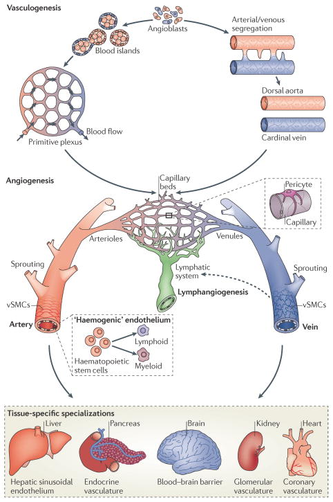
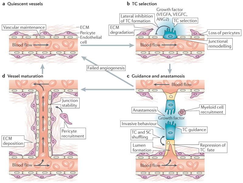
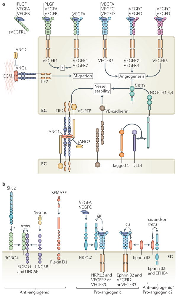
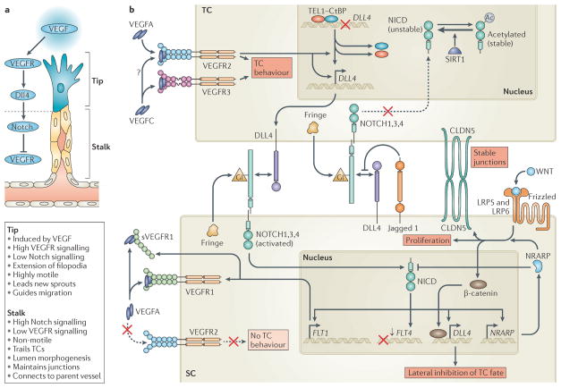
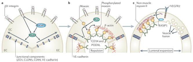
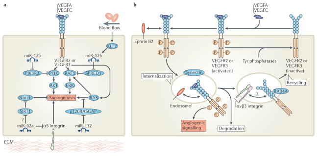
The vertebrate vasculature forms an extensive branched network of blood vessels that supplies tissues with nutrients and oxygen. During vascular development, coordinated control of endothelial cell behaviour at the levels of cell migration, proliferation, polarity, differentiation and cell-cell communication is critical for functional blood vessel morphogenesis. Recent data uncover elaborate transcriptional, post-transcriptional and post-translational mechanisms that fine-tune key signalling pathways (such as the vascular endothelial growth factor and Notch pathways) to control endothelial cell behaviour during blood vessel sprouting (angiogenesis). These emerging frameworks controlling angiogenesis provide unique insights into fundamental biological processes common to other systems, such as tissue branching morphogenesis, mechanotransduction and tubulogenesis.


Conflict of interest statement

Competing interests statement

The authors declare no competing financial interests.

Figures

Figure 1
Figure 1. Development of a functional vasculature from endothelial progenitor cells
Endothelial progenitors (angioblasts) differentiate from mesodermal cells during early vertebrate development. Once formed, angioblasts may acquire arterial (red) or venous (blue) fates and coalesce to generate the first embryonic blood vessels, the dorsal aorta and cardinal vein. In zebrafish, the coordinated sorting and segregation of arterial and venous angioblasts ensures the assembly of distinct dorsal aorta and cardinal vein vessels. Angioblasts also aggregate to form blood islands, which fuse and remodel in response to haemodynamic stimuli or inherent genetic factors to generate a primitive interlaced network of arterial and venous plexi. Following their vasculogenic assembly, angiogenic remodelling of the dorsal aorta, cardinal vein and vascular plexi creates a complex hierarchical network of arteries, arterioles, capillary beds, venules and veins. Subsequent recruitment of mural cells (pericytes and vascular smooth-muscle cells (vSMCs)) stabilizes nascent vessels and promotes vessel maturation. In addition, the sprouting of lymphatic endothelial cells from venous vessels (lymphangiogenesis) seeds the lymphatic system (indicated by a dotted arrow). Moreover, the emergence of haematopoietic stem cells from arterial ‘haemogenic’ endothelium gives rise to all myeloid and lymphoid blood cell lineages. Vessel diversity is further augmented by tissue-specific specializations that alter key properties, such as permeability, or modify endothelial cells to generate vascular networks with new molecular signatures,.
Figure 2
Figure 2. Cellular mechanisms of angiogenic sprouting
a | In the absence of pro-angiogenic stimuli, endothelial cells (ECs) are retained in a quiescent state. In addition, EC homeostasis is maintained by low-level autocrine vascular endothelial growth factor A (VEGFA) signalling. b | During angiogenesis, high levels of exogenous pro-angiogenic factors (such as VEGFA and VEGFC) and of VEGF receptor 2 (VEGFR2) or VEGFR3 signalling select ‘tip cells’ (TCs; blue) for sprouting. By contrast, Delta-like 4–Notch signalling laterally inhibits TC fate in adjacent ECs. TC sprouting behaviour is facilitated by the vascular endothelial cadherin-mediated loosening of EC–EC junctions, matrix metalloproteinase-mediated degradation of extracellular matrix (ECM) and the detachment of pericytes (purple). c | Invasive TC sprouting is guided by gradients of pro-angiogenic growth factors and various environmental guidance cues, such as semaphorins and ephrins. During sprout elongation, TCs are trailed by endothelial ‘stalk cells’ (SCs; yellow), which maintain connectivity with parental vessels and initiate partitioning-defective 3 (PAR3)-mediated vascular lumen morphogenesis. Expression of VEGFR1 and activation of Notch, Roundabout homologue 4 and WNT signalling in SCs repress TC behaviour to maintain the hierarchical organization of sprouting ECs. However, TCs and SCs may also shuffle and exchange positions during angiogenic sprouting. Upon contact with other vessels, TC behaviour is repressed and vessels fuse by the process of anastomosis, which is assisted by associated myeloid cells. d | Nascent perfused vessels are subsequently stabilized by the platelet-derived growth factor B-mediated recruitment of supporting pericytes, the strengthening of EC–EC contacts and the deposition of an ECM to re-establish a quiescent endothelial phenotype. ANG2, angiopoietin 2.
Figure 3
Figure 3. Key signalling pathways that control angiogenesis
a | General signalling pathways that control endothelial cell (EC) behaviour. Mammalian vascular endothelial growth factors (VEGFs) bind homodimers and heterodimers of three VEGF receptors (VEGFRs). Signalling via VEGFR2, VEGFR3 or VEGFR2 VEGFR3 heterodimers is pro-angiogenic. Proteolytic processing of VEGFC and VEGFD is required to permit their interaction with VEGFR2. VEGFA binding to VEGFR1 and the secreted VEGFR1 extracellular domain (soluble VEGFR1 (sVEGFR1)) acts as a sink for VEGFA that limits its availability to activate VEGFR2. The interaction of TIE2 receptor with matrix-associated angiopoietin 1 (ANG1) at EC–extracellular matrix (ECM) junctions induces migration. By contrast, at EC–EC junctions ANG1–TIE2 interactions promote quiescence upon trans-complex formation with TIE2 on adjacent cells. These complexes include vascular endothelial protein Tyr phosphatase (VE-PTP; also known as PTPRB) and activate distinct signalling pathways from those at cell–matrix contacts. ANG2 antagonizes ANG1 activity on TIE2 to destabilize vessels and aid angiogenic remodelling. Homophilic VE-cadherin interactions maintain EC–EC junctions. Delta-like 4 (DLL4)-mediated activation of Notch receptors represses angiogenic cell behaviour and promotes vessel stability upon the proteolytic release of the Notch intracellular domain (NICD). In certain contexts, Jagged 1 competes with DLL4 for Notch to decrease DLL4–Notch-mediated signalling. b | Known axon guidance receptors expressed in ECs. Roundabout homologue 4 (ROBO4)–uncoordinated 5 homologue B (UNC5B) interactions promote UNC5B-mediated inhibition of VEGFR signalling, block angiogenesis and maintain vessel integrity. Activation of ROBO4 by Slit 2 may also block VEGFR signalling but is controversial. Activation of UNC5B by netrins may also disrupt angiogenesis. Secreted class III semaphorins (such as semaphorin 3E (SEMA3E)) promote EC repulsion to perturb angiogenesis upon binding their receptor, plexin D1. By contrast, neuropilin 1 (NRP1) or NRP2 augment angiogenic EC behaviour on binding VEGFA or VEGFC and/or on interaction with VEGFR2 or VEGFR3. Activation of ephrin receptor B4 (EPHB4) upon interaction with its membrane-associated ligand, ephrin B2, may promote EC–EC repulsion or attraction in various cellular contexts and is essential for angiogenesis. Reverse EPHB4–ephrin B2 signalling may also play key parts in vascular development. Importantly, association of ephrin B2 with VEGFR2 or VEGFR3 promotes membrane internalization of the VEGFR and enhances angiogenic signalling. PLGF, placental growth factor.
Figure 4
Figure 4. Molecular mechanisms of endothelial tip cell selection
a | Sprouting endothelial cells are hierarchically organized into leading ‘tip cells’ (TCs) and trailing ‘stalk cells’ (SCs) that exhibit very distinct and specialized cell behaviours. TC formation is induced by vascular endothelial growth factor (VEGF) signalling, whereas Delta-like 4 (DLL4)–Notch signalling represses VEGF receptor (VEGFR) signalling and TC fate in SCs. b | VEGFA and VEGFC signalling via VEGFR2 and VEGFR3 induce the invasive and motile TC behaviour that drives angiogenesis. VEGFR2 activation induces DLL4 expression in TCs, which activates Notch on adjacent SCs. VEGF-mediated disruption of a repressive translocation ETS leukaemia (TEL) and carboxy-terminal-binding protein (CtBP) complex that binds theDLL4 promoter may partially account for the ability of VEGF to induceDLL4 expression. Notch signalling in SCs downregulates the expression of VEGFR3 (encoded by FLT4) and upregulates the expression of VEGFR1 (encoded by FLT1) and soluble VEGFR1 (sVEGFR1), which represses VEGFR2 function and blocks TC behaviour. Notch activation also inducesDLL4 expression in SCs to propagate the DLL4 Notch-mediated lateral inhibition of VEGFR2 and VEGFR3 along developing vessels. Notch-induced Notch-regulated ankyrin repeat-containing protein (NRARP) expression enhances WNT signalling in SCs, which maintains EC–EC junctions, promotes proliferation and may augment DLL4 expression via β-catenin. NRARP also promotes feedback inhibition of Notch signalling. Notch signalling in TCs is blocked by Jagged 1, which is expressed in SCs and impedes DLL4–Notch interactions on TCs when Notch is glycosylated (Gl) by Fringe family glycosyltransferases in the Golgi apparatus. Deacetylation of the Notch intracellular domain (NICD) by sirtuin 1 (SIRT1) may also negatively influence TC Notch signalling. CLDN5, claudin 5; LRP, low-density lipoprotein receptor-related protein.
Figure 5
Figure 5. Molecular mechanisms of lumen morphogenesis
a | Prior to lumen morphogenesis, blood vessels consist of coalesced cords of endothelial cells (ECs) that lack apicobasal polarity. β1 integrin–matrix interactions and RAS interacting protein 1 (RASIP1) establish EC apicobasal polarity in a partitioning defective 3 (PAR3)-mediated manner to promote the lateral redistribution of junctional components from the apical surface to the periphery of EC–EC contacts. b | Once EC apicobasal polarity is established, lumen formation is triggered, at least in part, by vascular endothelial cadherin (VE-cadherin)-mediated redistribution of CD34 and podocalyxin (PODXL) to the apical surface.β1 integrin may also promote the redistribution of PODXL to EC apical membranes. Subsequently, protein kinase C (PKC)-mediated phosphorylation and redistribution of moesin to PODXL-enriched apical EC membranes promotes the deposition of filamentous actin (F-actin). Furthermore, PODXL may initiate lumen formation by inducing the electrostatic repulsion of EC–EC apical surfaces. c | Lumenal expansion proceeds by a variety of mechanisms. For example, vascular endothelial growth factor receptor 2 (VEGFR2) signalling and activation of RHO-associated coiled-coil kinase (ROCK) may promote the association of non-muscle myosin II with apical F-actin to drive actomyosin-mediated cell shape changes. By contrast, RASIP1 may repress actomyosin contractility to fine-tune this response. Alternatively, or more likely in addition, directed exocytic vacuole trafficking and fusion of these vacuoles with the apical surface may also drive lumen expansion. ZO1, zonula occludens 1; CLDN5, claudin 5.
Figure 6
Figure 6. Fine-tuning angiogenic signals
a | Post-transcriptional modification of key angiogenic signalling pathways by microRNAs (miRNAs) modulates angiogenesis. Expression of miR-126 de-represses phosphoinositide 3-kinase (PI3K) and/or RAF1 signalling to promote vascular endothelial growth factor (VEGF)-induced angiogenesis. Blood flow also influences VEGF signalling by promoting Krüppel-like factor 2 (KLF2)-induced expression of miR-126 during angiogenic sprouting. Furthermore, endothelial cell expression of miR-132 enhances angiogenic signalling controlled by RAS small GTPases by reducing the expression of RAS-specific GTPase-activating protein p120 (p120RASGAP), a negative regulator of RAS. By contrast, miR-92a blocks angiogenesis by repressing pro-angiogenic α5 integrin protein expression. miR-92a may also activate Notch signalling to block VEGF-induced angiogenesis by reducing the expression of sirtuin 1 (SIRT1), which deacetylates the Notch intracellular domain to destabilize it. b | Post-translational modulation of VEGF receptor 2 (VEGFR2) or VEGFR3 membrane trafficking determines the duration and magnitude of VEGFA and VEGFC signalling. Synectin and ephrin B2-mediated membrane internalization protects phosphorylated active VEGFR from inactivation by cell-surface Tyr phosphatases. Furthermore, transport of VEGFR to intracellular endocytic compartments enhances pro-angiogenic signalling. Internalized VEGFR is subsequently either degraded or recycled to the cell surface for another round of activation. Hence, αvβ3 integrin and RAB4A-mediated recycling of VEGFR stabilizes receptor protein expression and enhances VEGF signalling. PIK3R2, PI3K regulatory subunit 2; SPRED1, Sprouty-related EVH1 domain-containing 1; ERK, extracellular signal-regulated kinase.

References

    1. Carmeliet P, Jain RK. Molecular mechanisms and clinical applications of angiogenesis. Nature. 2011;473:298–307. - PMC - PubMed
    1. Adams RH, Alitalo K. Molecular regulation of angiogenesis and lymphangiogenesis. Nature Rev Mol Cell Biol. 2007;8:464–478. - PubMed
    1. Rocha SF, Adams RH. Molecular differentiation and specialization of vascular beds. Angiogenesis. 2009;12:139–147. - PubMed
    1. Tammela T, Alitalo K. Lymphangiogenesis: molecular mechanisms and future promise. Cell. 2010;140:460–476. - PubMed
    1. Gaengel K, Genove G, Armulik A, Betsholtz C. Endothelial-mural cell signaling in vascular development and angiogenesis. Arterioscler Thromb Vasc Biol. 2009;29:630–638. - PubMed

Publication types

MeSH terms

Substances