Review
. 2011 Aug 23;12(9):605-20.
doi: 10.1038/nrm3173.

The predator becomes the prey: regulating the ubiquitin system by ubiquitylation and degradation

Affiliations
Review

The predator becomes the prey: regulating the ubiquitin system by ubiquitylation and degradation

Allan M Weissman et al. Nat Rev Mol Cell Biol. .

Erratum in

  • Nat Rev Mol Cell Biol. 2011 Oct;12(10):686

Abstract

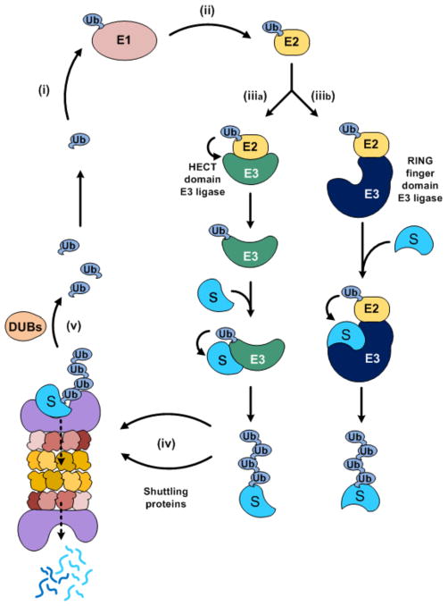
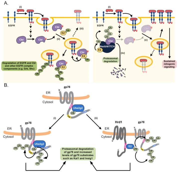
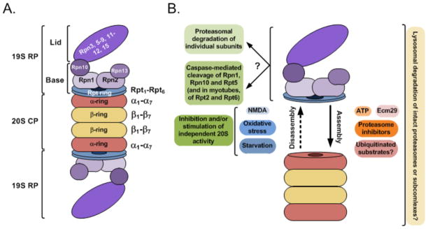
Ubiquitylation (also known as ubiquitination) regulates essentially all of the intracellular processes in eukaryotes through highly specific modification of numerous cellular proteins, which is often tightly regulated in a spatial and temporal manner. Although most often associated with proteasomal degradation, ubiquitylation frequently serves non-proteolytic functions. In light of its central roles in cellular regulation, it has not been surprising to find that many of the components of the ubiquitin system itself are regulated by ubiquitylation. This observation has broad implications for pathophysiology.


Figures

Figure 1
Figure 1. Schematic of the UPS
(i) Ubiquitin is activated by the ubiquitin-activating enzyme (E1), which uses ATP to form a high energy labile E1–thiol ester intermediate. (ii) The activated ubiquitin is transferred to a ubiquitin carrier protein (ubiquitin-conjugating enzyme; E2) to generate a similar thiol ester intermediate. (iiia) Activated ubiquitin can be transferred from the E2 to a HECT (Homologous to the E6-AP C-Terminus) domain ubiquitin protein-ligase (E3) to generate a third labile thiol ester intermediate. It is then transferred to the substrate (S), where, in most cases, a stable isopeptide bond between the activated carboxy terminal Gly76 of ubiquitin and an εNH2 group of an internal lysine residue in the substrate is formed. Additional ubiquitin moieties are then added to generate a polyubiquitin chain. (iiib) Alternatively, activated ubiquitin can be transferred directly from E2 to an internal lysine residue in a substrate that is bound to a RING finger ubiquitin ligase. Polyubiquitin chains linked through Lys48 of ubiquitin are characteristically associated with proteasomal degradation. However, linkages through several of the other seven lysines in ubiquitin have recently been shown to also target for proteasomal degradation. In some cases, ubiquitin is conjugated to the αNH2 group of the target substrate (reviewed in REF. 24), and there are a few examples of conjugation to internal substrate Thr, Ser, or Cys residues. Certain substrates are recognized by several ligases that act either under different conditions, sequentially or in parallel, recognizing different motifs in the substrate. Such a mechanism may be necessary to ascertain recognition and degradation of different domains in the substrate. (iv) Degradation of the ubiquitin-conjugated substrate to short peptides by the 26S proteasome. The binding of ubiquitinated proteins to the proteasome can be direct or mediated or enhanced by shuttle proteins (v) Most of the ubiquitin chain is disassembled by deubiquitinating enzymes (DUBs), the rest is probably degraded along with the substrate. Select substrates are processed rather than completely degraded by the 26S proteasome. This is due, at least in part, to unstructured sequences (for example, GlyAla repeats) that may inhibit entry into the proteasome, although additional mechanisms may be involved.
Figure 2
Figure 2. Modes of degradation of ubiquitin and ubiquitin-protein ligases
A. Ubiquitin can be degraded in three ways. (Aa) It can be degraded as a monomer following its polyubiquitylation (indicated in light blue). The process is probably slow, as the ubiquitin substrate molecule is short and lacks an unstructured tail that appears to be an important structural characteristic necessary for efficient proteasomal degradation. (Ab) Ubiquitin can be degraded with its conjugated substrate. The proximal part of the polyubiquitin chain is probably degraded along with the substrate, whereas the distal part is recycled by DUBs as free and reusable ubiquitin. (Ac) Ubiquitin with a C-terminal tail unstructured tail that is longer than 20 amino acid residues can be degraded efficiently by the proteasome. B. Modes of degradation of ubiquitin-protein ligases by ubiquitylation. (Ba) Degradation of a ligase mediated by self-ubiquitylation. Both RING and HECT domains-containing ligases can catalyze self-ubiquitylation, which generate polyubiquitin chains that target the proteins for proteasomal degradation. Self ubiquitylation is shown as occurring in cis (i.e. on the same molecule). However, as many E3s dimerize and may form higher order oligomers, ubiquitylation can also occur in trans as discussed in the text for Hrd1p. (Bb) Ubiquitylation of a ligase by a heterologous ligase. Whereas in the described cases of this mode of ubiquitylation the reaction is unidirectional (see Text and Figures 3 and 4), bidirectional ubiquitylation is also a possibility. (Bc) Hierarchical ubiquitylation of ligases. In this hypothetical scheme, one ligase targets several substrate ligases in a hierarchical manner. As has been described for several substrates of the ubiquitin system, one ligase substrate can be targeted by more than a single ligase (middle ligase in lower row and left ligase in middle row). The ligase at the top is targeted by self-ubiquitylation or by one of the ligases that it directly or indirectly regulates.
Figure 3
Figure 3. Targeting of Specific Ligases for Degradation by both Self- and Heterologous Ubiquitylation
A. Regulation of Cbl proteins and effects on signaling. Left hand side: (i) Binding of ligand induces dimerization and cross-phosphorylation of receptor tyrosine kinases (for example, EFGR – represented here) or receptor-associated tyrosine kinases as well as phosphorylation of other substrates, including other components of the signaling complex and Cbl proteins. The receptors together with other components of the signaling complex (not shown) are endocytosed and phosphorylated; Cbl proteins are recruited. (ii) Cbls mediate the ubiquitylation of receptors and associated proteins as well as self-ubiquitylation, resulting in targeting of receptors for lysosomal degradation and the destruction of Cbl molecules – whether this degradation is lysosomal or proteasomal has not been determined with certainty. (iii) The net result is decreased recycling of receptors to the cell surface and downregulation of signaling. Right hand side: (iv) Nedd4 family members target Cbl proteins for proteasomal degradation lowering the cellular levels of these E3s. (v) As a result ubiquitylation of activated receptor tyrosine kinase complexes is decreased. This results in attenuation of receptor down-regulation leading to increased mitogenic signaling. B. gp78 and ERAD. (i) gp78 is a polytopic RING finger E3 found in the ER that mediates self-ubiquitylation using the E2, Ube2g2. This activity requires an intact gp78 RING finger, the ubiquitin binding CUE domain (whose function in this process is not yet clear) and a highly specific binding site for Ube2g2 that is distinct from the RING finger. (ii) gp78 can additionally be targeted for ubiquitylation by a heterologous RING finger E3, Hrd1 (human Hrd1)/Synoviolin. This targeting is dependent on the Hrd1/Synoviolin RING finger but is independent of the gp78 RING finger. Both of these modes of degradation lead to proteasomal degradation of gp78 and consequently increased levels of gp78 substrates including Insig-1, and KAI1 (also known as CD82), which are regulators of cholesterol metabolism and a metastasis suppressor, respectively.
Figure 4
Figure 4. Regulation of specific ligases
A. Regulation of RING1B by ubiquitylation. (i) Self-ubiquitylation of RING1B generates ‘non-canonical’ - mixed (K6-, K-27-, and K-48-based) and multiply branched polyubiquitin chains that stimulate the monoubiquitinating activity of the ligase towards histone H2A, but do not target it for degradation. (ii) The HECT E3 E6-AP (and possibly an additional ligase) targets RING1B for proteasomal degradation by generating K48-based polyubiquitin chains. Both the self and the E6-AP-catalyzed ubiquitylations target the same lysine residues on RING1B, thus preventing opposing and wasteful activities - destruction of an active and needed enzyme, or untoward activation of an un-needed enzyme. (iii) USP7 de-ubiquitylates both the activating and the proteasome-targeting polyubiquitin chains. Its activity releases free RING1B, thus enabling inactivation, and at the same time stabilization of RING1B without destroying it. B. Regulation of Diap1. (i) Self-ubiquitylation of Diap1 generates K63-based polyubiquitin chains that attenuate the ligase activity towards its substrates such as caspases. (ii) Diap2 targets Diap1 for proteasomal degradation by generating K48-based polyubiquitin chains on Diap1. (iii) Following an apoptotic stimulus, Reaper, Hid and possibly Grim (which are known as the RHG family) induce accelerated self-ubiquitylation of Diap1, generating K48-based chains that target the protein for degradation. (iv) Morgue generates K48-based chains either independently, or in conjunction with the RHG family members. These chains target Diap1 for proteasomal degradation. C. Regulation of mammalian IAPs. In mammals both cellular IAP1 (cIAP1) and X chromosome-linked IAP (XIAP) can catalyze their self-ubiquitylation, generating K48-based chains that probably target them for proteasomal degradation. It is not known whether, like D. melanogaster IAP1 (Diap1), they can also generate non K48-based chains that serve non-proteolytic functions. Intact cIAP1 (or its RING finger domain alone) was shown to catalyze formation of K48-based chains on XIAP1 that target it for degradation. It is not known whether XIAP and/or another ligase can catalyze a similar reaction, targeting cIAP1 for degradation. The ligase SIAH1 can mediate generation of K48-based chains that target XIAP for degradation. This reaction is mediated by ARTS that probably allows for association of SIAH1 and XIAP. Similar to the RHG family members, Smac3 (and possibly Smac/DIABLO and ARTS) can induce accelerated auto-ubiquitylation of XIAP, which leads to its degradation. Solid arrows mark experimentally established ubiquitylations, broken arrows denote putative ubiquitylations. Ubiquitin-conjugating enzymes (E2s) are not represented graphically in this figure to reduce complexity, but should be assumed to be involved in all ubiquitylation reactions.
Figure 5
Figure 5. The 26S proteasome and its regulation by degradation
A. Structure of the 26S proteasome. The 26S proteasome is comprised of two sub-complexes - the 20S core catalytic particle (CP), and the 19S regulatory particle (RP). The substrate is first bound, probably via its polyubiquitin chain(s), to specific subunits in the 19S RP. It is then probably unfolded by ATPases residing in the base of the RP (Rpt ring), and inserted via an open gate in the α-ring of the 20S CP into the proteolytic chamber. The ATPases are probably also involved in opening the interlacing chains that close the gate into the α-ring of the CP, thus allowing entry of the unfolded substrate. The RP contains also DUB(s) that recycle ubiquitin and/or edit the polyubiquitin chain to enhance substrate binding to the RP and to recycle ubiquitin. A RP-associated ubiquitin ligase(s) may also function to adapt the chain length/type for optimal recognition by the RP. Proteolysis of the substrate is mediated by three pairs of proteolytically active β subunits inside the proteolytic chamber, which includes two adjacent β rings. B. Regulation of assembly and disassembly of the 26S proteasome, and degradation of its subunits. The entire proteasome or its sub-complexes are probably degraded by the lysosome via microautophagy. Different factors control assembly (ATP, Ecm29, ubiquitylated substrates, and proteasome inhibitors, for example) and disassembly of the proteasome to its RP and CP (for example, NMDA (N-methyl-D-aspartate) and different stresses, such as oxidative stress and starvation). The RP can be further disassembled into its individual subunits, which are probably degraded by proteasomes following ubiquitylation. For further reading on the proteasome structure and function, see REF. .

References

    1. Wilkinson KD. The discovery of the ubiquitin-dependent proteolysis. Proc Natl Acad Sci USA. 2005;102:15280–15282. - PMC - PubMed
    1. Shabek N, Iwai K, Ciechanover A. Ubiquitin is degraded by the ubiquitin system as a monomer and as part of its conjugated target. Biochem Biophys Res Commun. 2007;363:425–431. - PubMed
    1. Hershko A, Eytan E, Ciechanover A, Haas AL. Immunochemical analysis of the turnover of ubiquitin-protein conjugates in intact cells. Relationship to the breakdown of abnormal proteins. J Biol Chem. 1982;257:13964–13970. - PubMed
    2. The first description of the role of the ubiquitin proteolytic system in the degradation of proteins in nucleated cells. All prior studies describing the roles of the system were carried out using reticulocytes - the terminally differentiating red blood cell.

    1. Haas AL, Bright PM. The dynamics of ubiquitin pools within cultured human lung fibroblasts. J Biol Chem. 1987;262:345–351. - PubMed
    1. Patel MB, Majetschak M. Distribution and interrelationship of ubiquitin proteasome pathway component activities and ubiquitin pools in various porcine tissues. Physiol Res. 2007;56:341–350. - PubMed

Publication types

MeSH terms