. 2012 Apr 5;31(14):1845-58.
doi: 10.1038/onc.2011.368. Epub 2011 Aug 22.

FOXO3a represses VEGF expression through FOXM1-dependent and -independent mechanisms in breast cancer

Affiliations

FOXO3a represses VEGF expression through FOXM1-dependent and -independent mechanisms in breast cancer

C T Karadedou et al. Oncogene. .

Erratum in

Abstract

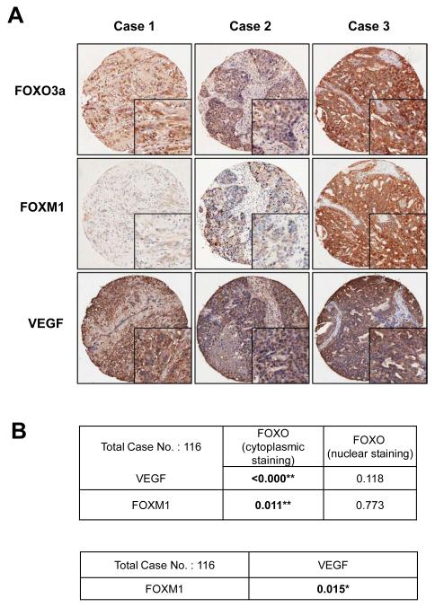
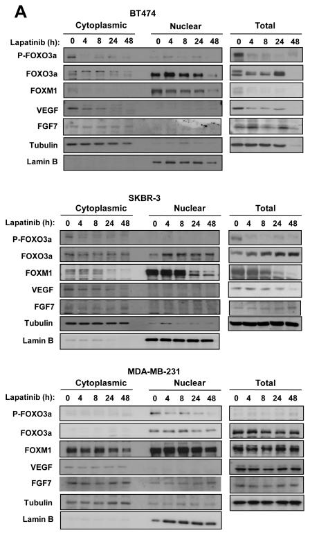
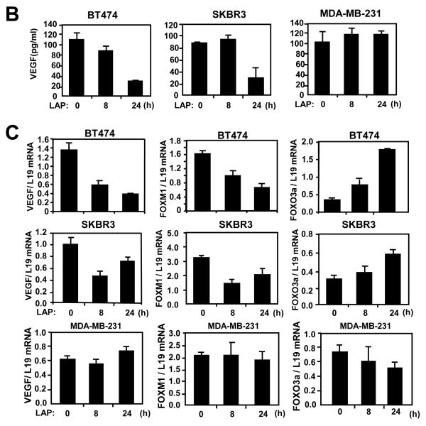
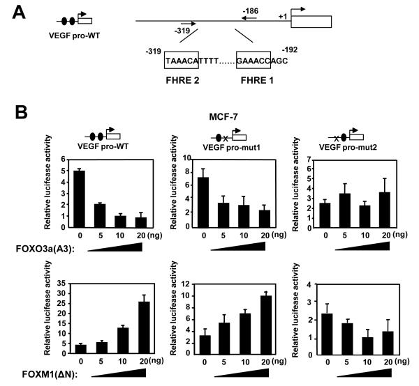
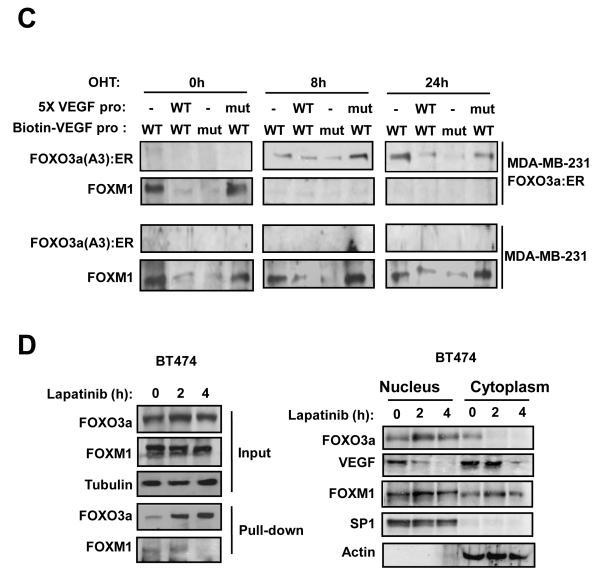
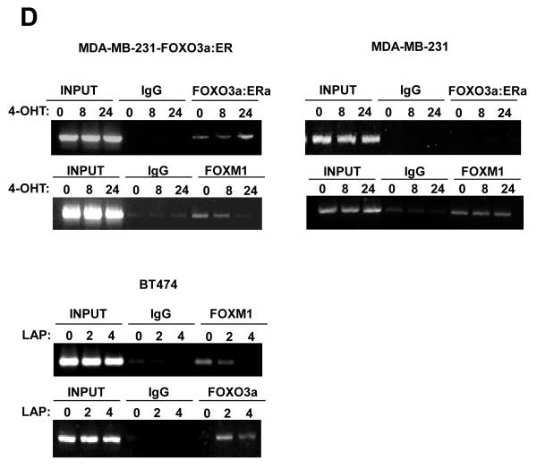
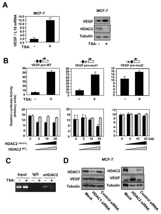
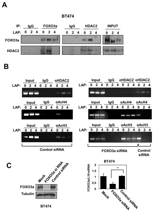
Vascular endothelial growth factor (VEGF) has a central role in breast cancer development and progression, but the mechanisms that control its expression are poorly understood. Breast cancer tissue microarrays revealed an inverse correlation between the Forkhead transcription factor Forkhead box class O (FOXO)3a and VEGF expression. Using the lapatinib-sensitive breast cancer cell lines BT474 and SKBR3 as model systems, we tested the possibility that VEGF expression is negatively regulated by FOXO3a. Lapatinib treatment of BT474 or SKBR3 cells resulted in nuclear translocation and activation of FOXO3a, followed by a reduction in VEGF expression. Transient transfection and inducible expression experiments showed that FOXO3a represses the proximal VEGF promoter, whereas another Forkhead member, FOXM1, induces VEGF expression. Chromatin immunoprecipitation and oligonucleotide pull-down assays showed that both FOXO3a and FOXM1 bind a consensus Forkhead response element (FHRE) in the VEGF promoter. Upon lapatinib stimulation, activated FOXO3a displaces FOXM1 bound to the FHRE before recruiting histone deacetylase 2 (HDAC2) to the promoter, leading to decreased histones H3 and H4 acetylation, and concomitant transcriptional inhibition of VEGF. These results show that FOXO3a-dependent repression of target genes in breast cancer cells, such as VEGF, involves competitive displacement of DNA-bound FOXM1 and active recruitment of transcriptional repressor complexes.


Figures

Figure 1
Figure 1. Representative expression patterns of FOXO3a, FOXM1 and VEGF in tissue microarray
One hundred and thirty-three cases of breast cancer diagnosed between the years 1992 to 2001 with clinical follow up data available were retrieved from the records of the Department of Pathology, Queen Mary Hospital of Hong Kong. The patients’ ages at diagnosis ranged from 30 to 90 years old, with a mean of 53 years. Histological sections of all cases were reviewed by the pathologist, the representative paraffin tumour blocks chosen as donor block for each case and the selected areas marked for construction of tissue microarray blocks. Tissue sections were deparaffinised, rehydrated and stained with a previously described primary polyclonal FOXO3a specific antibody (Nordigarden et al 2009, Rosivatz et al 2006) (diluted at 1:1400), the VEGF antibody (dilution 1:250) and FOXM1 (C-20, dilution 1:450, Santa Cruz). A total of 116 could be assessed and scored for FOXO3a, FOXM1 and VEGF expression using a scanscope (Scanscope Aperio Technologies, Inc, Vista, Calif) connected to a personal computer as described in figure S1. The expression pattern and subcellular localization were correlated with histological type, histological grade, clinical stage, estrogen and progestrogen receptor status, HER2 oncoprotein overexpression, lymph node metastasis and survival time (Fig. S2 and S3). Tumour tissue samples obtained from breast cancer patients that had been formalin-fixed and paraffin-embedded were immunohistochemically stained with FOXO3a, FOXM1 and VEGF antibodies using the streptavidin-biotin-peroxidase technique. Scoring was performed as described in Fig. S1. A) Three representative tumour cases showing corresponding FOXO3a FOXM1 and VEGF staining patterns (magnification: x 100; insets x400). The three cases 1, 2 and 3 represent low, medium and high FOXO3a cytoplasmic staining. Cases 1 and 2 also show nuclear FOXO3a staining and low FOXM1 and VEGF staining, while case 3 shows predominantly strong cytoplasmic FOXO3a staining, and strong FOXO3a and VEGF staining. B) Correlation analysis of FOXO3a, FOXM1 and VEGF staining in 116 breast carcinoma cases. The correlation between predominant nuclear/cytoplasmic FOXO3a expression with FOXM1 and VEGF expression and FOXM1 with VEGF expression was studied using Chi-Square Tests and was considered significant * at p≤0.05 and very significant ** at p≤0.01.
Figure 2
Figure 2. Expression of FOXO3a, FOXM1 and VEGF in response to lapatinib treatment in breast cancer cell lines
The lapatinib sensitive BT474 and SKBR-3 and resistant MDA-MB-231 cells were cultured in 10% FCS medium for 24 h before treatment with lapatinib. A) At times indicated, cells were collected and analysed for P-FOXO3a, total FOXO3a, FOXM1, VEGF, FGF7, LaminB and tubulin expression by western blotting of nuclear/cytoplamic (left panel) and total (right panel) lysates. B) VEGF concentrations in supernatants of the lapatinib-treated breast cancer cells were measured by a quantitative sandwich enzyme immunoassay according to the manufacturer’s protocol (Quantikine ELISA, R&D Systems, Abingdon, UK). The optical density was measured at 450nm using a Sunrise-Tecan plate reader (TECAN Ltd, Reading, UK) and VEGF concentrations normalised using standard curves. C) In parallel, VEGF mRNA levels of these lapatinib-treated breast cancer cells were also analysed by qRT-PCR and normalized to L19 RNA expression. Total RNA (2 μg) isolated using the RNeasy Mini kit (Qiagen, Crawley, UK) was reverse transcribed using the Superscript III reverse transcriptase and random primers (Invitrogen, Paisley, UK), and the resulting first strand cDNA was used as template in the real-time PCR. All experiments were performed in triplicate. The following gene-specific primer pairs were designed using the ABI Primer Express software: FOXM1-sense: 5′-TGCAGCTAGGGATGTGAATCTTC-3′ and FOXM1-antisense: 5′-GGAGCCCAGTCCATCAGAACT-3′; ERα-sense: 5′-CAGATGGTCAGTGCCTTGTTGG-3′ and ERα-antisense: 5′-CCAAGAGCAAGTTAGGAGCAAACAG-3′; L19-sense 5′-GCGGAAGGGTACAGCCAAT-3′ and L19-antisense 5′-GCAGCCGGCGCAAA-3′. Specificity of each primer was determined using NCBI BLAST module. Real time PCR was performed with ABI PRISM 7700 Sequence Detection System using SYBR Green Mastermix (Applied Biosystems, Brackley, UK). The RT-qPCR results shown are representative of 3 independent experiments. FOXM1 mRNA levels of these cells were also analysed by RT-qPCR, and normalized with L19 RNA expression.
Figure 3
Figure 3. FOXO3a represses the expression of FOXM1 and VEGF in the breast carcinoma cells MDA-MB-231 and MCF-7
A) MDA-MB-231-FOXO3a(A3):ER and MDA-MB-231 cells were treated with 200 nmol/L 4-OHT for the indicated times. Nuclear and cytoplasmic extracts were prepared at the times indicated, separated on polyacrylamide gels, and subjected to immunoblotting with specific antibodies. The expression levels of FOXO3a(A3):ER, FOXO3a, P-FOXO3a, FOXM1, VEGF, FGF7, tubulin and Lamin B1 were analyzed by Western blotting and the VEGF concentrations in supernatants of the 4-OHT-treated MDA-MB-231 cells cells were measured by the quantitative sandwich enzyme immunoassay as described in Fig. 2B. B) Total RNA was extracted from these cells and analyzed for FOXM1 and VEGF mRNA expression using RT-qPCR as described in the text and normalized to the level of L19 RNA. C) MCF-7 cells transiently transfected with the constitutively active FOXO3a(A3) or control vector were analysed for VEGF and FOXM1 expression by western blot and RT-qPCR analysis. D) MCF-7 cells transiently transfected with FOXO3a or control siRNA, or mock transfected were analysed by western blot using specific antibodies FOXO3a, FOXM1, VEGF and tubulin as indicated and by RT-qPCR for VEGF and FOXM1 mRNA expression. All data shown represent the averages of data from three experiments, and the error bars show the standard deviations. E) BT474 cells transiently transfected with control or FOXO3a siRNA, were treated with lapatinib for 0, 16, 24 and 48 h. Protein lysates were prepared at the times indicated and analyzed by western blot using specific antibodies P-HER2, HER-2, P-FOXO3a, FOXO3a, FOXM1, VEGF and tubulin as indicated.
Figure 4
Figure 4. FOXO3a represses and FOXM1 induces the transcriptional activity of the human VEGF gene through a FHRE consensus site proximal to the transcription start site
A) Effect of expression of FOXO3a and FOXM1 on VEGF promoter activity. Schematic representation of the VEGF-luciferase reporter construct, showing the consensus FHRE sequences. A 1741 bp VEGF promoter construct (positions −1,926 to −186 relative to the predominant 5′-transcription start site) was cloned into the XhoI and HindIII sites of the pGL3 basic vector (Promega, Southampton, United Kingdom). Putative forkhead site mutagenesis was performed using a Stratagene QuikChange site-directed mutagenesis kit with the oligonucleotides: Site1 (−178) (5′-ATCCCTCTTCTTTTTTCTTGGGCATTTTTTTTTAAAACTGTATTGT-3′), and Site2 (−319) (5′- TTGCTCTACTTCCCCGGGTCACTGTGGATTTTGGGGGCCAGCAGA-3′). B) MCF-7 cells were transiently transfected with 20 ng of either the wild-type, (VEGF pro-WT), mutant FHRE1 (VEGF pro-mut1), or mutant FHRE2 (VEGF pro-mut2) VEGF promoter/reporter and 0, 5, 10 or 20 ng of either the constitutively active FOXO3a(A3) or FOXM1(ΔN) expression vector. Cells were harvested 24 h after transfection and assayed for luciferase activity. All relative luciferase activity values are corrected for cotransfected Renilla activity. All data shown represent the averages of data from three independent experiments, and the error bars show the standard deviations. C) MDA-MB-231-FOXO3a(A3):ER and MDA-MB-231 cells were treated with 200 nmol/L 4-OHT for the indicated times. Nuclear extracts prepared were incubated with biotinylated wild-type or mutant FHRE2 oligonucleotides in the presence or absence of 5x molar excess of non-biotinylated wild-type or mutant FHRE2 oligonucleotides. Proteins binding to the biotinylated oligonucleotides were pulled-down using streptavidine agarose beads and analysed by western blot using specific antibodies as indicated. D) The nuclear and cytoplasmic extracts prepared from BT474 cells treated with lapatinib for 0, 2 and 4 h were western blotted for proteins indicated (right panel). The nuclear extracts from the lapatinib-treated cells were also examined by pull-down assays using biotinylated wild-type or mutant FHRE2 oligonucleotides as described above. E) Chromatin immunoprecipitation (ChIP) analysis of the human VEGF promoter. MDA-MB-231-FOXO3a(A3):ER, MDA-MB-231 and BT474 cells described above were used for ChIP assays using IgG, anti-FOXO3a and anti-FOXM1 antibodies as indicated. After crosslink reversal, the co-immunoprecipitated DNA was amplified by PCR using primers amplifying the VEGF FHRE2 containing region (−351/−186) and resolved in 2% agarose gel. Representative data from three independent experiments are shown.
Figure 5
Figure 5. Effects of HDAC2 overexpression and depletion on the expression of VEGF in MCF-7 cells
A) MCF-7 cells treated with vehicle or 100 nM trichostatin A (TSA; Sigma, UK) for 24 h were harvested for RT-qPCR and western blot analysis for VEGF expression. B) MCF-7 cells transiently transfected with 20 ng of either the wild-type, (VEGF pro-WT), mutant FHRE1 (VEGF pro-mut1), or mutant FHRE2 (VEGF pro-mut2) VEGF promoter/reporter were either untreated or treated with 100 nM TSA, or co-tranfected with 0, 5, 10 or 20 ng of either the wild-type or constitutively active HDAC2 expression vector. The transfected cells were then harvested for luciferase assays after 24 h. C) MCF-7 cells untreated or treated with TSA for 24 h were analysed for HDAC2 binding on the VEGF promoter by ChIP assays as described. D) MCF-7 cells were transiently transfected with control and smart pool siRNA against either HDAC1 or HDAC2 and analysed by western blotting for protein expression as indicated.
Figure 6
Figure 6. OXO3a recruits HDAC2 to the VEGF promoter in response to lapatinib in BT474 cells
A) Cell extracts prepared from BT474 cells 0, 2 and 4 h after treatment with lapatinib were immunoprecipitated (IP) with antibodies against FOXO3a and HDAC2 or an IgG control antibody. The precipitated complexes and the inputs were examined for FOXO3a and HDAC2 expression. B) BT474 cells were transiently transfected with smart pool siRNA against FOXO3a or control siRNA pool. Twenty-four h afterwards, the transfected BT474 cells were treated with lapatinib for 0, 2, or 4 h and then analysed for HDAC2, acetylated histone H3 and H4 binding on the VEGF promoter by ChIP assays as described. C) Western blot and RT-qPCR analyses were performed as described to demonstrate effective and specific FOXO3a knock-down.

Similar articles

Cited by

References

    1. Arden KC. FOXO animal models reveal a variety of diverse roles for FOXO transcription factors. Oncogene. 2008;27:2345–2350. - PubMed
    1. Bernstein BE, Kamal M, Lindblad-Toh K, Bekiranov S, Bailey DK, Huebert DJ, et al. Genomic maps and comparative analysis of histone modifications in human and mouse. Cell. 2005;120:169–181. - PubMed
    1. Burgering BM. A brief introduction to FOXOlogy. Oncogene. 2008;27:2258–2262. - PubMed
    1. Calnan DR, Brunet A. The FoxO code. Oncogene. 2008;27:2276–2288. - PubMed
    1. Ciardiello F. Epidermal growth factor receptor inhibitors in cancer treatment. Future Oncol. 2005;1:221–234. - PubMed

Publication types