. 2011 Jun 26;3(6):53-62.
doi: 10.4252/wjsc.v3.i6.53.

Short and long term fate of human AMSC subcutaneously injected in mice

Affiliations

Short and long term fate of human AMSC subcutaneously injected in mice

Pilar López-Iglesias et al. World J Stem Cells. .

Abstract

Aim: To study the ability of human adipose-derived mesenchymal stem cells (AMSCs) to survive over the short and long term, their biodistribution and their biosafety in vivo in tumor-prone environments.

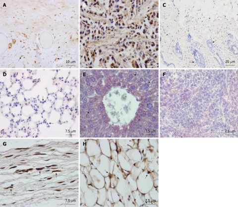
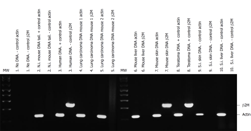
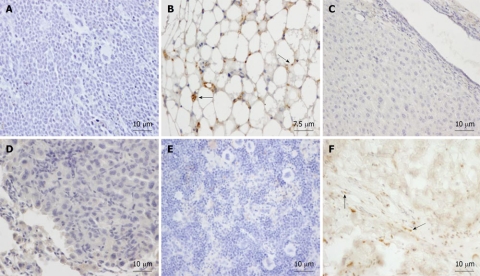
Methods: We subcutaneously injected human AMSCs from different human donors into immunodeficient SCID mice over both short- (2 and 4 mo) and long- (17 mo) term in young, and aged tumor-prone mice. Presence of human cells was studied by immunohistochemistry and polymerase chain reaction analysis in all organs of injected mice.

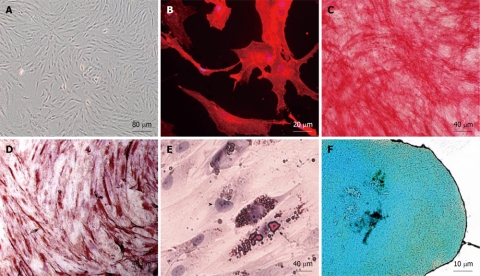
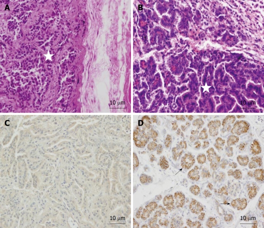
Results: Subcutaneously injected AMSCs did not form teratomas at any time point. They did not migrate but remained at the site of injection regardless of animal age, and did not fuse with host cells in any organ examined. AMSCs survived in vivo for at least 17 mo after injection, and differentiated into fibroblasts of the subdermic connective tissue and into mature adipocytes of fat tissue, exclusively at the site of injection.

Conclusion: Our results support the assertion that AMSC may be safe candidates for therapy when injected subcutaneously because of their long term inability to form teratomas.

Keywords: Adipose-derived stem cells; Biosafety; Cell therapy; Cell transplant; Mesenchymal stem cells; SCID mice; Teratoma.


Figures

Figure 1
Figure 1
In vitro characterization of human adipose-derived mesenchymal stem cell. A: Phase contrast of a primary subconfluent culture showing typical adherent fibroblast-like morphology; B: Anti-CD90 immunofluorescence (in red) of a human adipose-derived mesenchymal stem cell (hAMSC) culture; C: Assessment of in vitro differentiation potential of hAMSC by osteogenic differentiation, demonstrated by alkaline phosphatase activity in red; D: Assessment of in vitro differentiation potential of hAMSC by myogenic differentiation, demonstrated by anti-sarcomeric-α-actin in brown (arrow); E: Assessment of in vitro differentiation potential of hAMSC by adipogenic differentiation, demonstrated by Oil red. Arrow: fat drops in a labeled differentiated cell. Arrowhead: Nucleus of an undifferentiated cell; F: Assessment of in vitro differentiation potential of hAMSC by chondrogenic differentiation, demonstrated by Alcian blue.
Figure 2
Figure 2
Tissue sections from NOD/SCID mice transplanted subcutaneously with human adipose-derived mesenchymal stem cells. Mice were sacrificed at two mo and organs were formalin-fixed and paraffin-embedded. Sections were immunostained with monoclonal anti-human nuclear ribonucleoprotein antibody and counterstained with Harris hematoxylin. A: Human Positive control of the antibody, showing intense nuclear immunostaining in the testicular albuginea in brown. Arrows: human labeled fibroblasts; B: Positive control. SCID subepidermal teratoma showing positive nuclear staining for anti-human ribonucleoprotein. Arrows: human labeled teratoma cells; C: Saline injected SCID showing no staining for anti-human ribonucleoprotein in the skin at the site of injection. Arrows: mouse unlabelled cells of the hair follicles; D: Lung of an adipose-derived mesenchymal stem cell (AMSC) injected SCID mouse at 2 mo showing no human AMSC (hAMSC). Arrows: mouse unlabelled cells; E: Liver of an AMSC injected SCID mouse at 2 mo showing no hAMSC. Arrows: mouse unlabelled cells; F: Spleen of an AMSC injected SCID mouse at 2 mo showing no hAMSC. Arrows: mouse unlabelled lymphocytes; G: Subcutaneous connective tissue of an AMSC injected mouse at the site of injection, showing positive nuclear staining in the fibroblasts in brown (arrows); H: Subdermic adipose tissue of an AMSC injected mouse at the site of injection, showing positive nuclear staining in the adipocytes (arrows).
Figure 3
Figure 3
Detection of human DNA from adipose-derived mesenchymal stem cells in NOD/SCID transplanted mice tissues. Human and mouse DNA were amplified by polymerase chain reaction with specific human β2 microglobulin primers (235 bp, β2M) and with actin primers that recognize mouse and human DNA (122 bp, actin) as mentioned in Materials and Methods. Each pair of lines shows the same DNA amplified with actin and with β2M primers. The following tissues of short-term transplanted mice (4 mo after cell transplantation) are shown: (1) Negative (-) control. No DNA in the polymerase chain reaction with actin or β2M primers; (2) Tail DNA from non-injected (N.i.) mouse tail DNA with actin (+ control of mouse DNA) or β2M primers (- control of human DNA); (3) Positive control. Human corneal tissue (human DNA) with actin or β2M primers; (4) Lung carcinoma DNA of a human adipose-derived mesenchymal stem cell (hAMSC) injected mouse 1 with actin or β2M primers; (5) Lung carcinoma DNA of another hAMSC injected mouse (2) with actin or β2M primers; (6) Liver DNA of a hAMSC injected mouse with actin or β2M primers; (7) Skin DNA of a hAMSC injected mouse with actin or β2M primers; (8) Teratoma DNA of NTERA-2 cells injected mouse with actin or β2M primers (positive (+) control for both sets of primers); (9) Negative control. Skin DNA from the saline injected (S.I.) mouse with actin or β2M primers; and (10) Negative control. Liver DNA from the saline injected mouse with actin or β2M primers.
Figure 4
Figure 4
Histopathological findings in paraffin embedded tissue sections of human adipose-derived mesenchymal stem cell transplanted 1-year-old SCID mice after 4 mo. A: NTERA-2 cell derived subepidermal teratoma from positive control transplanted mouse. HE. Star: Teratoma cells; B: Lung papillary carcinoma found in NOD/SCID mice transplanted subcutaneously with human adipose-derived mesenchymal stem cells after 4 mo. HE. Star: Carcinoma cells; C: Immunohistochemical analysis of lung papillary carcinoma showing no staining for anti-human mitochondria; D: Positive control of anti-human mitochondria in human pancreas showing strong immunoreaction in brown (arrows).
Figure 5
Figure 5
Paraffin-embedded tissue sections from NOD/SCID mice transplanted subcutaneously with human adipose-derived mesenchymal stem cells after 17 mo. Sections were immunostained with monoclonal anti-human mitochondria antibody (A-D) or anti-human ribonucleoprotein antibody (E, F) and then were counterstained with hematoxylin. A: Saline injected SCID showing no staining for anti-human mitochondria in spleen. Negative control; B: SCID subcutaneous adipose tissue section at the site of injection at 17 mo showing staining for anti human mitochondria in the adipocytes in brown (arrows); C: Liver of a human adipose-derived mesenchymal stem cell (AMSC) SCID injected mouse at 17 mo showing no human AMSC (hAMSC); D: Lung of a human AMSC SCID injected mouse at 17 mo showing no hAMSCs; E: Spleen of a human AMSC SCID injected mouse at 17 mo showing no hAMSCs; F: SCID subcutaneous dermal tissue section at the site of injection at 17 mo showing staining for anti human ribonucleoprotein in the fibroblasts in brown (arrows).

References

    1. Barry FP, Murphy JM. Mesenchymal stem cells: clinical applications and biological characterization. Int J Biochem Cell Biol. 2004;36:568–584. - PubMed
    1. Jiang Y, Jahagirdar BN, Reinhardt RL, Schwartz RE, Keene CD, Ortiz-Gonzalez XR, Reyes M, Lenvik T, Lund T, Blackstad M, et al. Pluripotency of mesenchymal stem cells derived from adult marrow. Nature. 2002;418:41–49. - PubMed
    1. Gronthos S, Mankani M, Brahim J, Robey PG, Shi S. Postnatal human dental pulp stem cells (DPSCs) in vitro and in vivo. Proc Natl Acad Sci U S A. 2000;97:13625–13630. - PMC - PubMed
    1. Zuk PA, Zhu M, Ashjian P, De Ugarte DA, Huang JI, Mizuno H, Alfonso ZC, Fraser JK, Benhaim P, Hedrick MH. Human adipose tissue is a source of multipotent stem cells. Mol Biol Cell. 2002;13:4279–4295. - PMC - PubMed
    1. Zuk PA, Zhu M, Mizuno H, Huang J, Futrell JW, Katz AJ, Benhaim P, Lorenz HP, Hedrick MH. Multilineage cells from human adipose tissue: implications for cell-based therapies. Tissue Eng. 2001;7:211–228. - PubMed