. 2011 Jul 28;3(7):182-7.
doi: 10.4329/wjr.v3.i7.182.

Endovascular management in abdominal visceral arterial aneurysms: A pictorial essay

Affiliations

Endovascular management in abdominal visceral arterial aneurysms: A pictorial essay

Manisha Jana et al. World J Radiol. .

Abstract

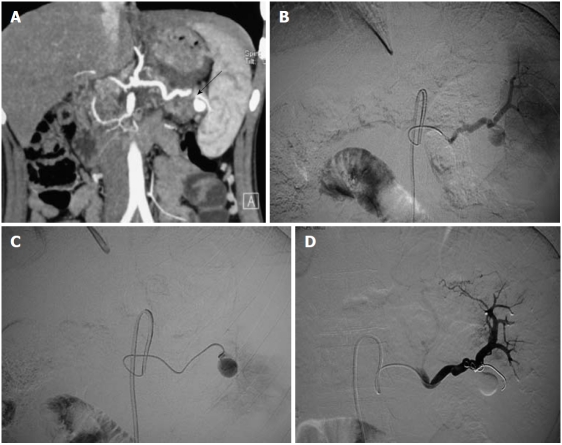
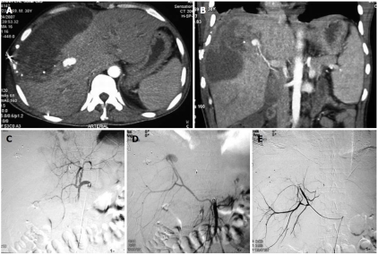
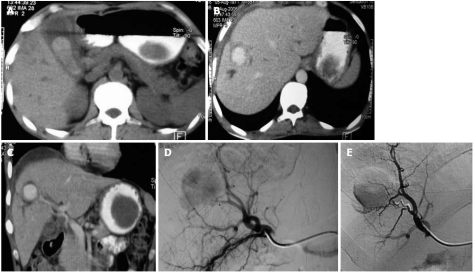
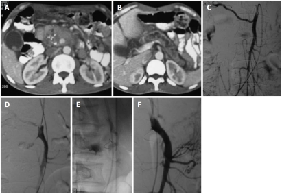
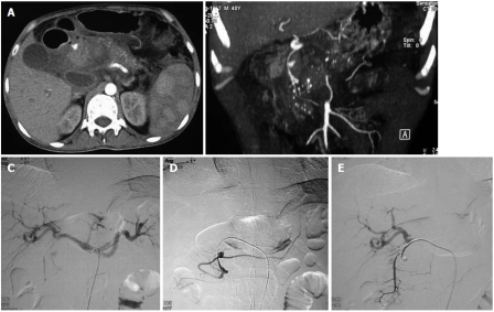
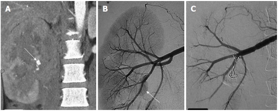
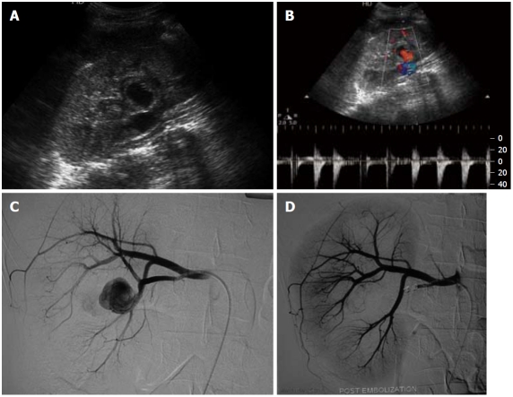
Visceral artery aneurysms (VAAs) include aneurysms of the splanchnic circulation and those of the renal artery. Their diagnosis is clinically important because of the associated high mortality and potential complications. Splenic, superior mesenteric, gastroduodenal, hepatic and renal arteries are some of the common arteries affected by VAAs. Though surgical resection and anastomosis still remains the treatment of choice in some of the cases, especially cases involving the proximal arteries, increasingly endovascular treatment is being used for more distal vessels. We present a pictorial review of various intra-abdominal VAAs and their endovascular management.

Keywords: Coil embolization; Endovascular management; Imaging; Pseudoaneurysm; Visceral arterial aneurysm.


Figures

Figure 1
Figure 1
Splenic artery aneurysm in a 21-year-old asymptomatic female. A: Coronal maximum intensity projection image of computed tomography angiogram reveals a pseudoaneurysm from the distal splenic artery at the hilum with a narrow neck (arrow); B, C: Digital subtraction angiography images reveal the pseudoaneurysm; D: After selective catheterization, the aneurysm was treated by coiling which resulted in complete occlusion in the aneurysm sac; visualized in the post-procedure image.
Figure 2
Figure 2
Traumatic pseudoaneurysm of a replaced right hepatic artery. A 38-year-old male presented with hypotension and falling hematocrit after a road traffic accident. A, B: Axial contrast-enhanced computed tomography of the abdomen (A) and coronal MIP image (B) revealed a large pseudoaneurysm (arrows) in the right lobe of the liver and a large hematoma in the liver parenchyma extending to the subcapsular location; C: DSA image after selective catheterization of the celiac trunk failed to reveal any aneurysm; D: Selective superior mesenteric artery (SMA) catheterization revealed a replaced right hepatic artery arising from the SMA and a pseudoaneurysm arising from it; E: Treated with coil embolization.
Figure 3
Figure 3
Traumatic right hepatic artery aneurysm. A 45-year-old male presented 2 mo after a road traffic accident with melena. A: Abdominal non-contrast computerized tomography revealed a hyperdense hematoma in the gall bladder lumen; B, C: Abdominal contrast-enhanced computed tomography axial (B) and coronal reformatted images (C) revealed a large pseudoaneurysm of the right hepatic artery (arrows); D: Selective catheter angiogram of the hepatic artery revealed the large pseudoaneurysm; E: Successful treatment by coil embolization (e).
Figure 4
Figure 4
Coil embolization in an superior mesenteric artery aneurysm in a patient with chronic calcific pancreatitis. A, B: Axial contrast-enhanced computed tomography of the abdomen revealed dilated main pancreatic duct and coarse calcification in the head of the pancreas, a large partially thrombosed pseudoaneurysm was apparent as a contrast filled globular structure in the head; C, D: Selective abdominal angiography of the superior mesenteric artery revealed the jet of injected contrast into the pseudoaneurysm cavity (D); E, F: Coil embolization was performed to occlude the neck (E) which resulted in complete occlusion and non-filling of the aneurysm (F).
Figure 5
Figure 5
Endovascular management of a gastroduodenal artery aneurysm secondary to chronic calcific pancreatitis. A, B: Axial image (A) and coronal reformatted image (B) of a computed tomography angiogram reveal features of acute on chronic calcific pancreatitis and a small saccular pseudoaneurysm of the gastroduodenal artery; C, D: Selective angiogram of the celiac axis (C) and gastroduodenal artery (D) reveal the filling of the aneurysm from the main trunk; E: Coil embolization was performed to fill the aneurysm cavity and cause complete occlusion.
Figure 6
Figure 6
Endovascular coil embolization of a left gastric artery causing upper gastrointestinal hemorrhage. A: Selective angiogram of the left gastric artery shows the large saccular aneurysm; B: Selective angiogram of the celiac axis after coil embolization revealed complete non-opacification of the left gastric artery aneurysm.
Figure 7
Figure 7
Traumatic right renal pseudoaneurysm. A: Coronal maximum intensity projection computed tomography angiographic image reveals a large renal midpolar laceration and a pseudoaneurysm (arrow); B: Selective angiogram of the main right renal artery reveals a pseudoaneurysm (arrow) arising from one of the posterior branches; C: Successfully occluded using coil.
Figure 8
Figure 8
Traumatic right renal artery pseudoaneurysm. A, B: Grey scale ultrasound (A) and Doppler ultrasound (B) of the right kidney reveal a pseudoaneurysm in the midpolar region; C, D: Selective angiogram (C) of the right renal artery shows the pseudoaneurysm which was successfully coil-embolized (D).

References

    1. Shanley CJ, Shah NL, Messina LM. Common splanchnic artery aneurysms: splenic, hepatic, and celiac. Ann Vasc Surg. 1996;10:315–322. - PubMed
    1. Liu Q, Lu JP, Wang F, Wang L, Jin AG, Wang J, Tian JM. Visceral artery aneurysms: evaluation using 3D contrast-enhanced MR angiography. AJR Am J Roentgenol. 2008;191:826–833. - PubMed
    1. Hallett JW. Splenic artery aneurysms. Semin Vasc Surg. 1995;8:321–326. - PubMed
    1. Gabelmann A, Görich J, Merkle EM. Endovascular treatment of visceral artery aneurysms. J Endovasc Ther. 2002;9:38–47. - PubMed
    1. O'Driscoll D, Olliff SP, Olliff JF. Hepatic artery aneurysm. Br J Radiol. 1999;72:1018–1025. - PubMed