Shedding light on class-specific wiring: development of intrinsically photosensitive retinal ganglion cell circuitry
- PMID: 21861091
- PMCID: PMC3230729
- DOI: 10.1007/s12035-011-8199-8
Shedding light on class-specific wiring: development of intrinsically photosensitive retinal ganglion cell circuitry
Abstract
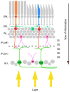
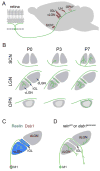
Neural circuits associated with retinal ganglion cells have long been used as models for investigating the mechanisms that govern circuit development and function. Similar to neurons in the brain, retinal ganglion cells are subdivided into distinct classes based upon their morphology, physiology, and patterns of connectivity. Newly developed transgenic tools in which individual classes of retinal ganglion cells are labeled with reporter proteins have recently provided a method to study the development of their class-specific circuitry. Here, we examine a single class of intrinsically photosensitive retinal ganglion cells and discuss their class-specific circuitry, as well as the cellular and molecular mechanisms that govern assembly of this circuitry.
Figures


Similar articles
-
Tracer coupling of intrinsically photosensitive retinal ganglion cells to amacrine cells in the mouse retina.J Comp Neurol. 2010 Dec 1;518(23):4813-24. doi: 10.1002/cne.22490. J Comp Neurol. 2010. PMID: 20963830 Free PMC article.
-
Photoreceptive Ganglion Cells Drive Circuits for Local Inhibition in the Mouse Retina.J Neurosci. 2021 Feb 17;41(7):1489-1504. doi: 10.1523/JNEUROSCI.0674-20.2020. Epub 2021 Jan 4. J Neurosci. 2021. PMID: 33397711 Free PMC article.
-
Genetic address book for retinal cell types.Nat Neurosci. 2009 Sep;12(9):1197-204. doi: 10.1038/nn.2370. Epub 2009 Aug 2. Nat Neurosci. 2009. PMID: 19648912
-
[Viral and Electrophysiological Approaches for Elucidating the Structure and Function of Retinal Circuits].Yakugaku Zasshi. 2018;138(5):669-678. doi: 10.1248/yakushi.17-00200-1. Yakugaku Zasshi. 2018. PMID: 29710012 Review. Japanese.
-
The injury resistant ability of melanopsin-expressing intrinsically photosensitive retinal ganglion cells.Neuroscience. 2015 Jan 22;284:845-853. doi: 10.1016/j.neuroscience.2014.11.002. Epub 2014 Nov 10. Neuroscience. 2015. PMID: 25446359 Free PMC article. Review.
Cited by
-
Central melanopsin projections in the diurnal rodent, Arvicanthis niloticus.Front Neuroanat. 2015 Jul 14;9:93. doi: 10.3389/fnana.2015.00093. eCollection 2015. Front Neuroanat. 2015. PMID: 26236201 Free PMC article.
-
Contributions of VLDLR and LRP8 in the establishment of retinogeniculate projections.Neural Dev. 2013 Jun 13;8:11. doi: 10.1186/1749-8104-8-11. Neural Dev. 2013. PMID: 23758727 Free PMC article.
-
Distribution and development of molecularly distinct perineuronal nets in visual thalamus.J Neurochem. 2018 Dec;147(5):626-646. doi: 10.1111/jnc.14614. Epub 2018 Nov 26. J Neurochem. 2018. PMID: 30326149 Free PMC article.
-
Nuclei-specific differences in nerve terminal distribution, morphology, and development in mouse visual thalamus.Neural Dev. 2014 Jul 10;9:16. doi: 10.1186/1749-8104-9-16. Neural Dev. 2014. PMID: 25011644 Free PMC article.
-
Aging Alters Daily and Regional Calretinin Neuronal Expression in the Rat Non-image Forming Visual Thalamus.Front Aging Neurosci. 2021 Feb 24;13:613305. doi: 10.3389/fnagi.2021.613305. eCollection 2021. Front Aging Neurosci. 2021. PMID: 33716710 Free PMC article.
References
-
- Masland RH. The fundamental plan of the retina. Nat Neurosci. 2001;4:877–886. - PubMed
-
- Masland RH. Neuronal diversity in the retina. Curr Opin Neurobiol. 2001;11:431–436. - PubMed
-
- Muscat L, Huberman AD, Jordan CL, Morin LP. Crossed and uncrossed retinal projections to the hamster circadian system. J Comp Neurol. 2003;466:513–524. - PubMed
Publication types
MeSH terms
Grants and funding
LinkOut - more resources
Full Text Sources

