

Sensory deprivation differentially impacts the dendritic development of pyramidal versus non-pyramidal neurons in layer 6 of mouse barrel cortex
- PMID: 21861159
- PMCID: PMC3737741
- DOI: 10.1007/s00429-011-0342-9
Sensory deprivation differentially impacts the dendritic development of pyramidal versus non-pyramidal neurons in layer 6 of mouse barrel cortex
Abstract
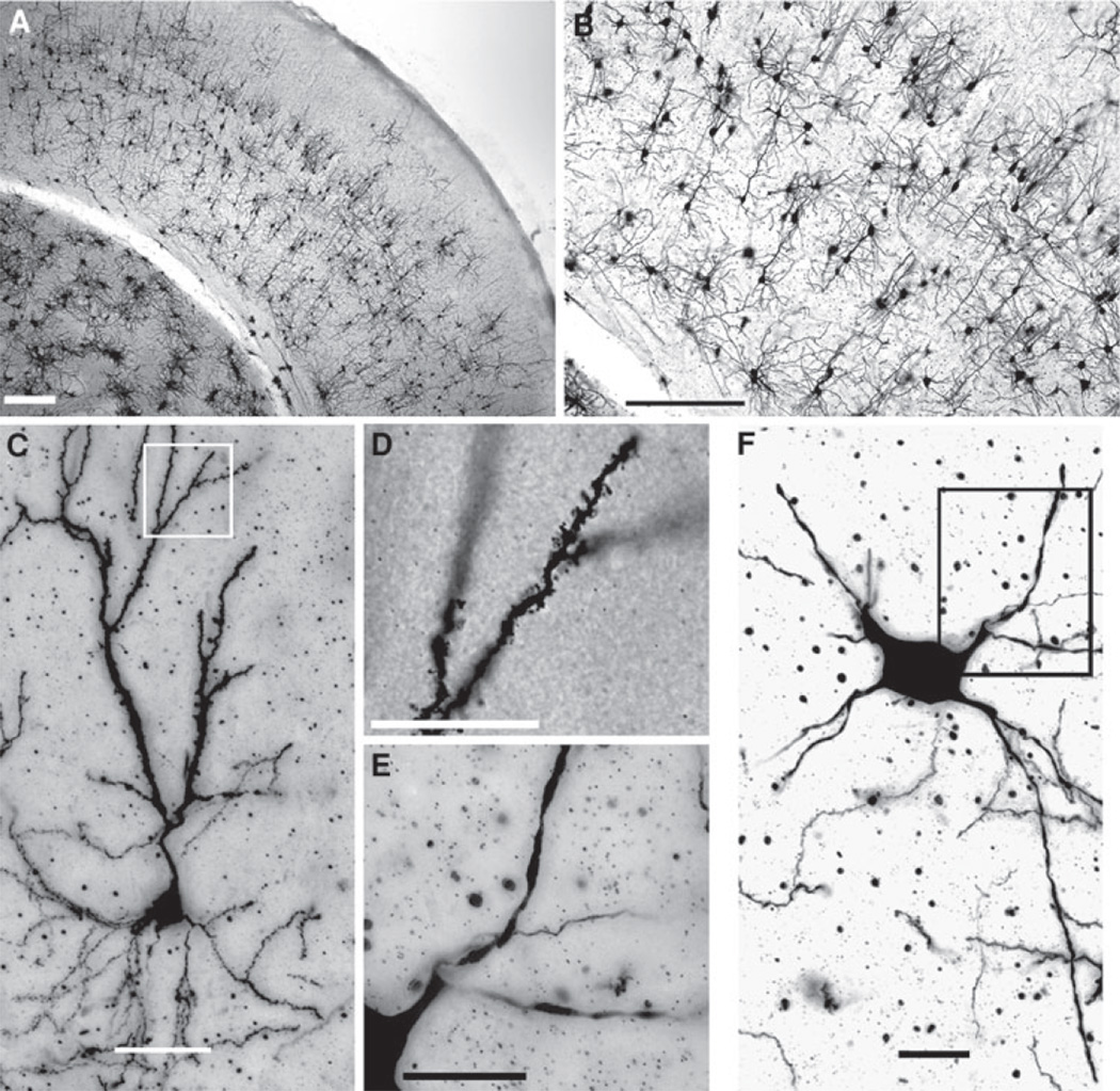
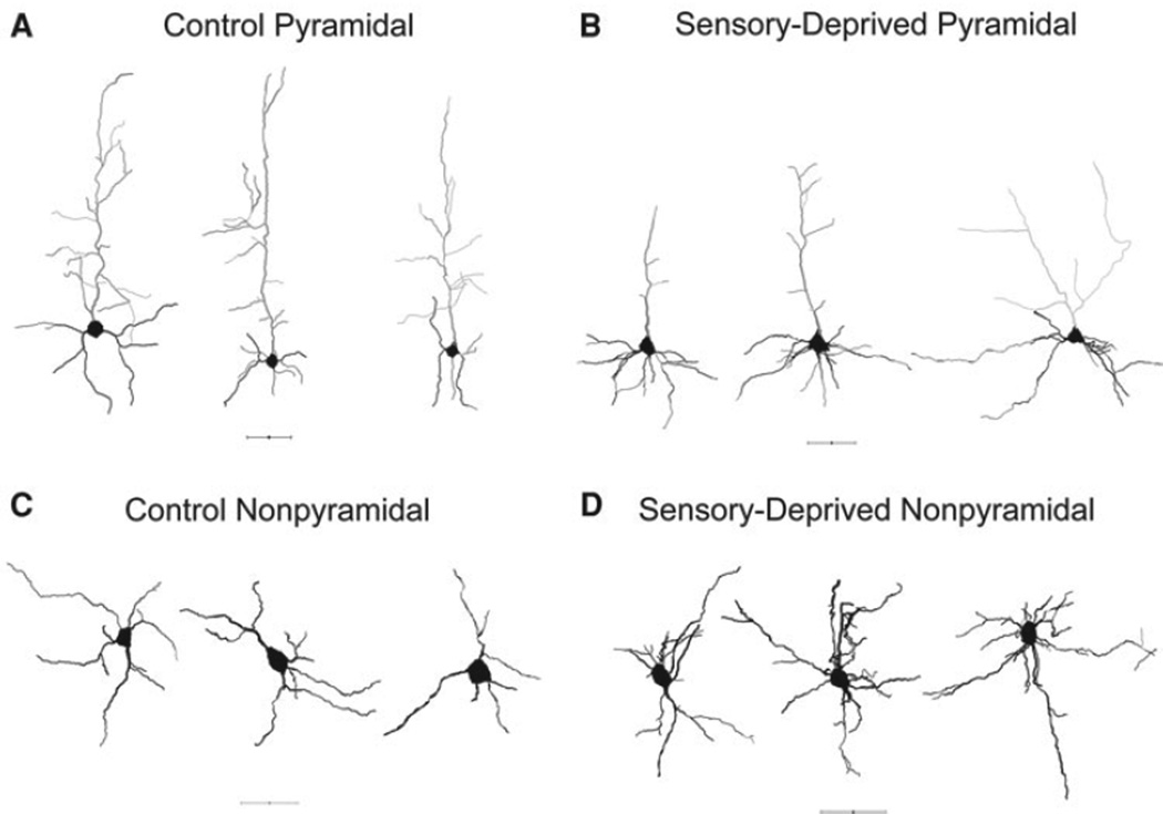
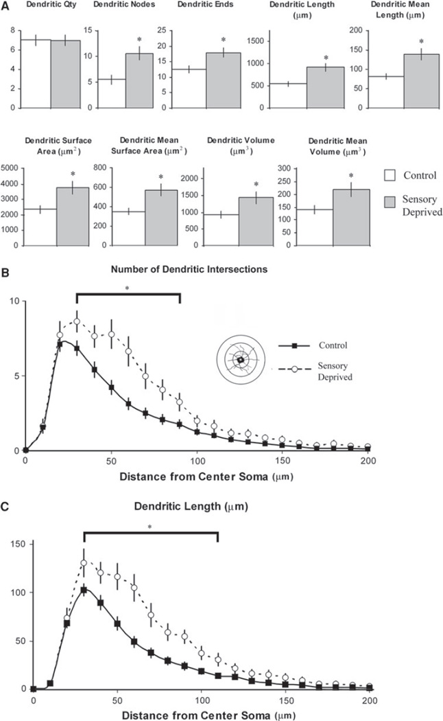
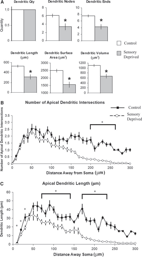
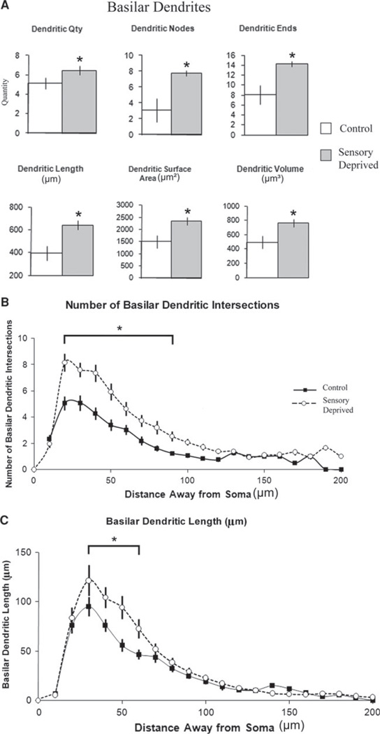
Early postnatal sensory experience can have profound impacts on the structure and function of cortical circuits affecting behavior. Using the mouse whisker-to-barrel system we chronically deprived animals of normal sensory experience by bilaterally trimming their whiskers every other day from birth for the first postnatal month. Brain tissue was then processed for Golgi staining and neurons in layer 6 of barrel cortex were reconstructed in three dimensions. Dendritic and somatic parameters were compared between sensory-deprived and normal sensory experience groups. Results demonstrated that layer 6 non-pyramidal neurons in the chronically deprived group showed an expansion of their dendritic arbors. The pyramidal cells responded to sensory deprivation with increased somatic size and basilar dendritic arborization but overall decreased apical dendritic parameters. In sum, sensory deprivation impacted on the neuronal architecture of pyramidal and non-pyramidal neurons in layer 6, which may provide a substrate for observed physiological and behavioral changes resulting from whisker trimming.
Figures
Similar articles
-
Loss of sensory input increases the intrinsic excitability of layer 5 pyramidal neurons in rat barrel cortex.J Physiol. 2009 Nov 1;587(Pt 21):5107-19. doi: 10.1113/jphysiol.2009.180943. Epub 2009 Sep 7. J Physiol. 2009. PMID: 19736297 Free PMC article.
-
Bilateral whisker trimming during early postnatal life impairs dendritic spine development in the mouse somatosensory barrel cortex.J Comp Neurol. 2010 May 15;518(10):1711-23. doi: 10.1002/cne.22297. J Comp Neurol. 2010. PMID: 20235164
-
Layer 4 pyramidal neurons exhibit robust dendritic spine plasticity in vivo after input deprivation.J Neurosci. 2015 May 6;35(18):7287-94. doi: 10.1523/JNEUROSCI.5215-14.2015. J Neurosci. 2015. PMID: 25948276 Free PMC article.
-
Experience-dependent changes in function and anatomy of adult barrel cortex.Exp Brain Res. 1998 Nov;123(1-2):110-6. doi: 10.1007/s002210050551. Exp Brain Res. 1998. PMID: 9835399 Review.
-
Mapping functional connectivity in barrel-related columns reveals layer- and cell type-specific microcircuits.Brain Struct Funct. 2007 Sep;212(2):107-19. doi: 10.1007/s00429-007-0147-z. Epub 2007 Jun 26. Brain Struct Funct. 2007. PMID: 17717691 Review.
Cited by
-
Development and sensory experience dependent regulation of microglia in barrel cortex.J Comp Neurol. 2020 Mar 1;528(4):559-573. doi: 10.1002/cne.24771. Epub 2019 Oct 18. J Comp Neurol. 2020. PMID: 31502243 Free PMC article.
-
Oligodendrocyte- and Neuron-Specific Nogo-A Restrict Dendritic Branching and Spine Density in the Adult Mouse Motor Cortex.Cereb Cortex. 2018 Jun 1;28(6):2109-2117. doi: 10.1093/cercor/bhx116. Cereb Cortex. 2018. PMID: 28505229 Free PMC article.
-
mGluR5 knockout mice display increased dendritic spine densities.Neurosci Lett. 2012 Aug 22;524(1):65-8. doi: 10.1016/j.neulet.2012.07.014. Epub 2012 Jul 20. Neurosci Lett. 2012. PMID: 22819970 Free PMC article.
-
The impact of development and sensory deprivation on dendritic protrusions in the mouse barrel cortex.Cereb Cortex. 2015 Jun;25(6):1638-53. doi: 10.1093/cercor/bht415. Epub 2014 Jan 9. Cereb Cortex. 2015. PMID: 24408954 Free PMC article.
-
High-throughput morphometric and transcriptomic profiling uncovers composition of naïve and sensory-deprived cortical cholinergic VIP/CHAT neurons.EMBO J. 2023 Jan 4;42(1):e110565. doi: 10.15252/embj.2021110565. Epub 2022 Nov 15. EMBO J. 2023. PMID: 36377476 Free PMC article.
References
-
- Bestman J, Santos da Silva J, Cline HT. Dendrite development. In: Stuart G, Spruston N, Häusser M, editors. Dendrites. NY: Oxford University Press; 2008. pp. 35–67.
-
- Briner A, De Roo M, Dayer A, Muller D, Kiss JZ, Vutskits L. Bilateral whisker trimming during early postnatal life impairs dendritic spine development in the mouse somatosensory barrel cortex. J Comp Neurol. 2010;518(10):1711–1723. - PubMed
-
- Buonomano DV, Merzenich MM. Cortical plasticity: from synapses to maps. Ann Rev Neurosci. 1998;21:149–186. - PubMed
Publication types
MeSH terms
Grants and funding
LinkOut - more resources
Full Text Sources
