Fine structure of the sensilla and immunolocalisation of odorant binding proteins in the cerci of the migratory locust, Locusta migratoria
- PMID: 21861654
- PMCID: PMC3281470
- DOI: 10.1673/031.011.5001
Fine structure of the sensilla and immunolocalisation of odorant binding proteins in the cerci of the migratory locust, Locusta migratoria
Abstract
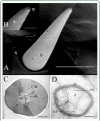
Using light and electron microscopy (both scanning and transmission), we observed the presence of sensilla chaetica and hairs on the cerci of the migratory locust, Locusta migratoria L. (Orthoptera: Acrididae). Based on their fine structures, three types of sensilla chaetica were identified: long, medium, and short. Males presented significantly more numbers of medium and short sensilla chaetica than females (p<0.05). The other hairs can also be distinguished as long and short. Sensilla chaetica were mainly located on the distal parts of the cerci, while hairs were mostly found on the proximal parts. Several dendritic branches, enveloped by a dendritic sheath, are present in the lymph cavity of the sensilla chaetica. Long, medium, and short sensilla chaetica contain five, four and three dendrites, respectively. In contrast, no dendritic structure was observed in the cavity of the hairs. By immunocytochemistry experiments only odorant-binding protein 2 from L. migratoria (LmigOBP2) and chemosensory protein class I from the desert locust, Schistocerca gregaria Forsskål (SgreCSPI) strongly stained the outer lymph of sensilla chaetica of the cerci. The other two types of hairs were never labeled. The results indicate that the cerci might be involved in contact chemoreception processes.
Figures


Similar articles
-
The chemosensilla on tarsi of Locusta migratoria (Orthoptera: Acrididae): distribution, ultrastructure, expression of chemosensory proteins.J Morphol. 2009 Nov;270(11):1356-63. doi: 10.1002/jmor.10763. J Morphol. 2009. PMID: 19530095
-
Developmental expression of odorant-binding proteins and chemosensory proteins in the embryos of Locusta migratoria.Arch Insect Biochem Physiol. 2009 Jun;71(2):105-15. doi: 10.1002/arch.20303. Arch Insect Biochem Physiol. 2009. PMID: 19408312
-
Expression of odorant-binding and chemosensory proteins and spatial map of chemosensilla on labial palps of Locusta migratoria (Orthoptera: Acrididae).Arthropod Struct Dev. 2006 Mar;35(1):47-56. doi: 10.1016/j.asd.2005.11.001. Arthropod Struct Dev. 2006. PMID: 18089057
-
Expression and immunolocalisation of odorant-binding and chemosensory proteins in locusts.Cell Mol Life Sci. 2005 May;62(10):1156-66. doi: 10.1007/s00018-005-5014-6. Cell Mol Life Sci. 2005. PMID: 15928808 Free PMC article.
-
Chemical Ecology and Olfaction in Short-Horned Grasshoppers (Orthoptera: Acrididae).J Chem Ecol. 2022 Feb;48(2):121-140. doi: 10.1007/s10886-021-01333-3. Epub 2022 Jan 10. J Chem Ecol. 2022. PMID: 35001201 Review.
Cited by
-
Dietary diversification and variations in the number of labrum sensilla in grasshoppers: which came first?J Biosci. 2013 Jun;38(2):339-49. doi: 10.1007/s12038-013-9325-8. J Biosci. 2013. PMID: 23660669
References
-
- Angeli S, Ceron F, Scaloni A, Monti M, Monteforti G, Minnocci A, Petacchi R, Pelosi P. Purification, structural characterization, cloning and immunocytochemical localization of chemoreception proteins from Schistocerca gregaria. European Journal of Biochemistry. 1999;262:745–754. - PubMed
-
- Ban LP, Scaloni A, Brandazza A, Angeli S, Zhang L, Yan YH, Pelosi P. Chemosensory proteins of Locusta migratoria. Insect Molecular Biology. 2003b;12(2):125–134. - PubMed
-
- Blaney WM. Electrophysiological responses of the terminal sensilla on the maxillary palps of Locusta migratoria (L.) to some electrolytes and non-electrolytes. Journal of Experimental Biology. 1974;60:275–293. - PubMed
-
- Blaney WM, Chapman RF. The fine structure of the terminal sensilla of the maxillary palps of Schistocerca gregaria (Forskål) (Orthoptera, Acrididae). Zeitschrift Fur Zellforschung Und Mikroskopische Anatomie. 1969;99:74–97. - PubMed
MeSH terms
Substances
LinkOut - more resources
Full Text Sources

