. 2012 May 20;21(8):1250-63.
doi: 10.1089/scd.2010.0578. Epub 2011 Oct 19.

A model of early human embryonic stem cell differentiation reveals inter- and intracellular changes on transition to squamous epithelium

Affiliations

A model of early human embryonic stem cell differentiation reveals inter- and intracellular changes on transition to squamous epithelium

Vasiliy Galat et al. Stem Cells Dev. .

Abstract

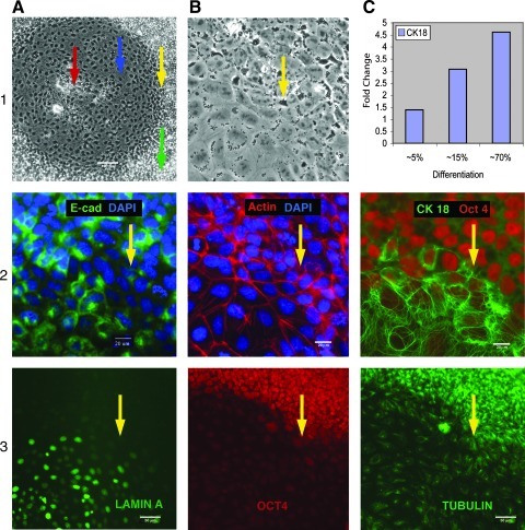
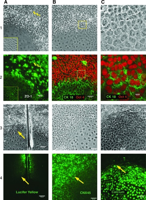
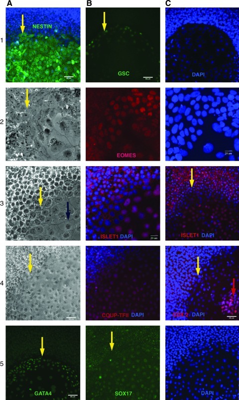
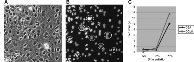
The molecular events leading to human embryonic stem cell (hESC) differentiation are the subject of considerable scrutiny. Here, we characterize an in vitro model that permits analysis of the earliest steps in the transition of hESC colonies to squamous epithelium on basic fibroblast growth factor withdrawal. A set of markers (GSC, CK18, Gata4, Eomes, and Sox17) point to a mesendodermal nature of the epithelial cells with subsequent commitment to definitive endoderm (Sox17, Cdx2, nestin, and Islet1). We assayed alterations in the transcriptome in parallel with the distribution of immunohistochemical markers. Our results indicate that the alterations of tight junctions in pluripotent culture precede the beginning of differentiation. We defined this cell population as "specified," as it is committed toward differentiation. The transitional zone between "specified" pluripotent and differentiated cells displays significant up-regulation of keratin-18 (CK18) along with a decrease in the functional activity of gap junctions and the down-regulation of 2 gap junction proteins, connexin 43 (Cx43) and connexin 45 (Cx45), which is coincidental with substantial elevation of intracellular Ca2+ levels. These findings reveal a set of cellular changes that may represent the earliest markers of in vitro hESC transition to an epithelial phenotype, before the induction of gene expression networks that guide hESC differentiation. Moreover, we hypothesize that these events may be common during the primary steps of hESC commitment to functionally varied epithelial tissue derivatives of different embryological origins.


Figures

FIG. 1.
FIG. 1.
A general outline of differentiation is shown (1A). Blue arrow indicates a typical area of flat cells of squamous epithelium. Red arrow—the central clusters of advanced differentiation. Green arrow—undifferentiated cells, yellow arrow—the cells of transitional zone. E-Cadherin (CM14) staining showing that adherens junction was present in hESCs and compromised in transitional zone, but became pronounced on differentiation (2A). F-actin cytoskeleton (H14) extended around the inner surface of the cell membrane typical of the epithelial cells “circumferential actin belt.” Cytoplasmic stress fibers became apparent when cells acquired epithelial morphology (1, 2B). CK18 (H14) is drastically up-regulated on epithelial differentiation; overlay with Oct4 (2C). RT-PCR analysis (H9) of CK18 expression showing gradual increase of the proportion of epithelial cells on differentiation progression (1C). Expression of Lamin A/C emerging in differentiating areas (3A) concurrent with Oct 4 down-regulation (3B). A developing network of tubulin in the differentiated cells (H14) is shown (3C). hESC, human embryonic stem cell; RT-PCR, reverse transcription–polymerase chain reaction.
FIG. 2.
FIG. 2.
There was considerable expression of ZO-1 (1, 2A) in undifferentiated cultures (CM14); however, expression was lost near the border of differentiated areas (yellow lightening). ZO-1 was associated with tight junction as differentiation progressed to a defined epithelial phenotype (yellow inset). Oct4 (1, 2B) was down-regulated in differentiated area (H14); however, it remained intense within the zone of surrounding cells (pretransitional zone) where ZO-1 expression was altered [compare with (1, 2A)]. Higher magnification (1, 2C) of the cells in a transitional zone (yellow inset in 2B) with up-regulation of CK18 on the background of pluripotent morphology of hESCs with high levels of Oct4 expression (2C), although some evidence of cell “flattening” and nuclear elongation became apparent (1C). Scrape loading/dye transfer assay (CM14) (3, 4A). LY actively diffused through the functional gap junctions of the pluripotent cells; however, the LY diffusion was undetectable between the differentiated cells. The colonies (CM14) underwent a progressive loss of Connexin 45 expression in the differentiated cells (3, 4B). The green fluorescence (Fluo4) revealed significant elevation of intracellular Ca2+ level in the differentiating parts of the hESC colonies (H14) (3, 4C). The arrows indicate the border between the transitional zone and the differentiated cells. LY, lucifer yellow.
FIG. 3.
FIG. 3.
Lineage marker analysis. Marker of progenitor cells Nestin is up-regulated in epithelial cells (DSV2) overlay with DAPI is shown (1A). Mesendodermal markers Goosecoid protein (H14) (1B,C) and Eomes (DSV2) (2A–C) are expressed in the border with undifferentiated cells and are downregulated when the differentiation progresses, and cells acquire epithelial morphology. Markers of primitive and definitive endoderm: Gata4 (H14) is more pronounced in the regions adjusted to pluripotent cells (4A), COUP-TFII (H9) is expressed throughout the differentiated colony (4A, B) in a similar manner as Sox 17 (DSV2) (5B, C). Markers of definitive endoderm Islet 1 (3AC) have a similar pattern and are expressed in the nuclei of a vast majority of differentiated epithelia cells (DSV2). Islet 1, however, also accumulated in the perimembrane space but not in the nuclei of undifferentiated cells. Cdx2 is primarily detected in the central (older) parts of differentiated colonies (H14) overlaid with DAPI (4C). Blue arrows indicate a typical area of flat squamous epithelial cells. Red arrows indicate central clusters of advanced differentiation. Yellow arrows indicate cells of transitional zones.
FIG. 4.
FIG. 4.
Multinucleated cells (H14) typical for syncytiotrophoblasts are seen in the epithelial culture (A, B). RT-PCR analysis (H9) showed notable up-regulation of CG alpha and GCM1 when differentiation spreads to 70% of culture aria (C). GCM1, glial cells missing homolog 1.
FIG. 5.
FIG. 5.
(A) Unsupervised clustering based on the gene content; (B) Unsupervised clustering based on the distribution of top ranked gene ontogeny (GO) processes in enrichment analysis. (C) Differentially expressed genes, which passed 1.5-FC thresholds for a corresponding pair of FGF2+\FGF2− cell lines. (Up-, down-regulated in the FGF2− cell lines). FGF2, basic fibroblast growth factor.

Similar articles

Cited by

References

    1. Murry C. Keller G. Differentiation of embryonic stem cells to clinically relevant populations: lessons from embryonic development. Cell. 2008;132:661–680. - PubMed
    1. Cai J. Chen J. Liu Y. Miura T. Luo Y. Loring JF. Freed WJ. Rao MS. Zeng X. Assessing self-renewal and differentiation in human embryonic stem cell lines. Stem cells. 2006;24:516–530. - PMC - PubMed
    1. Bhattacharya B. Cai J. Luo Y. Miura T. Mejido J. Brimble SN. Zeng X. Schulz TC. Rao MS. Puri RK. Comparison of the gene expression profile of undifferentiated human embryonic stem cell lines and differentiating embryoid bodies. BMC Dev Biol. 2005;5:22–38. - PMC - PubMed
    1. Vallier L. Alexander M. Pedersen RA. Activin/Nodal and FGF pathways cooperate to maintain pluripotency of human embryonic stem cells. J Cell Sci. 2005;118:4495–4509. - PubMed
    1. Eiselleova L. Matulka K. Kriz V. Kunova M. Schmidtova Z. Neradil J. Tichy B. Dvorakova D. Pospisilova S. Hampl A. Dvorak P. A complex role for FGF-2 in self-renewal, survival, and adhesion of human embryonic stem cells. Stem Cells. 2009;27:1847–1857. - PMC - PubMed

Publication types

MeSH terms