Review
. 2011 Aug 2;13(4):231.
doi: 10.1186/ar3358.

Giant cell arteritis: immune and vascular aging as disease risk factors

Affiliations
Review

Giant cell arteritis: immune and vascular aging as disease risk factors

Shalini V Mohan et al. Arthritis Res Ther. .

Abstract

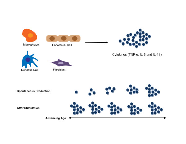
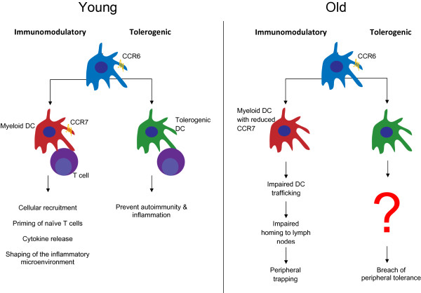
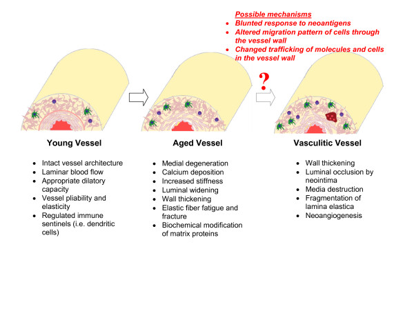
Susceptibility for giant cell arteritis increases with chronological age, in parallel with age-related restructuring of the immune system and age-induced remodeling of the vascular wall. Immunosenescence results in shrinkage of the naïve T-cell pool, contraction of T-cell diversity, and impairment of innate immunity. Aging of immunocompetent cells forces the host to take alternative routes for protective immunity and confers risk for pathogenic immunity that causes chronic inflammatory tissue damage. Dwindling immunocompetence is particularly relevant as the aging host is forced to cope with an ever growing infectious load. Immunosenescence coincides with vascular aging during which the arterial wall undergoes dramatic structural changes and medium and large arteries lose their pliability and elasticity. On the molecular level, elastic fibers deteriorate and matrix proteins accumulate biochemical modifications. Thus, the aging process impacts the two major biologic systems that liaise to promote giant cell arteritis; the immune system and the vessel wall niche.


Figures

Figure 1
Figure 1
Excessive production of pro-inflammatory cytokines in immune aging. The aging process results in an increase in basal cytokine production by macrophages, dendritic cells, endothelial cells and fibroblasts. Whereas young cells require stimulation to secrete cytokines and thus are closely controlled, old cells release cytokines spontaneously. The elevation in spontaneous cytokine production in the absence of specific stimulation results in a pro-inflammatory environment in which tissues are constantly 'primed' by cytokines. Chronic cytokine triggering may result in the generation of neoantigens and may contribute to the exhaustion of reserve pools supplying T cells and B cells. There is a corresponding decline in the ability of the immune system to mount a robust response as chronic cytokine exposure may alter the rheostat of cellular responsiveness.
Figure 2
Figure 2
Aging of dendritic cells. Aging affects both of the major functions of dendritic cells (DCs); stimulation and amplification of immunity and induction of peripheral tolerance. DCs are responsible for initiating and directing the immune response to antigens and play an integral role in the prevention of autoimmunity and unrestrained inflammation. Aged DCs are unable to efficiently prime and recruit T cells, migrate to peripheral lymph structures or maintain tolerance to self. They become trapped in peripheral tissues, perpetuating the immune response and can cause unresolved smoldering inflammation and subsequent tissue damage.
Figure 3
Figure 3
Age-related changes in vessel wall structure that enhance susceptibility to vasculitis. The vessel wall is prone to age-related changes that accumulate over a lifetime. Vascular stiffening results from biochemical changes of the cellular and extracellular components comprising the wall. Aged vessels typically lose elasticity and pliability. Elastic fibers/membranes show fatigue and fracture and matrix proteins accumulate biochemical modififications. Cell numbers and cell mobility through the wall layers change. The overall effect is a thinning of the medial layer, luminal widening and expansion of the intimal layer. Calcium deposition in the wall occurs frequently and may indicate foci of tissue injury. The 'old' artery provides a distinct microenvironment that potentially increases the risk for the homing and the persistence of pro-inflammatory immune cells and supplies a novel spectrum of neoantigens.

Similar articles

Cited by

References

    1. Weyand CM, Goronzy JJ. Medium- and large-vessel vasculitis. N Engl J Med. 2003;349:160–169. doi: 10.1056/NEJMra022694. - DOI - PubMed
    1. Ma-Krupa W, Jeon MS, Spoerl S, Tedder TF, Goronzy JJ, Weyand CM. Activation of arterial wall dendritic cells and breakdown of self-tolerance in giant cell arteritis. J Exp Med. 2004;199:173–183. doi: 10.1084/jem.20030850. - DOI - PMC - PubMed
    1. Han JW, Shimada K, Ma-Krupa W, Johnson TL, Nerem RM, Goronzy JJ, Weyand CM. Vessel wall-embedded dendritic cells induce T-cell autoreactivity and initiate vascular inflammation. Circ Res. 2008;102:546–553. doi: 10.1161/CIRCRESAHA.107.161653. - DOI - PubMed
    1. Pryshchep O, Ma-Krupa W, Younge BR, Goronzy JJ, Weyand CM. Vessel specific Toll-like receptor profiles in human medium and large arteries. Circulation. 2008;118:1276–1284. doi: 10.1161/CIRCULATIONAHA.108.789172. - DOI - PMC - PubMed
    1. Weyand CM, Younge BR, Goronzy JJ. T cells in arteritis and atherosclerosis. Curr Opin Lipidol. 2008;19:469–477. doi: 10.1097/MOL.0b013e32830bfdc2. - DOI - PMC - PubMed