. 2011 Aug 23;6(1):61.
doi: 10.1186/1750-1326-6-61.

A glycine zipper motif mediates the formation of toxic β-amyloid oligomers in vitro and in vivo

Affiliations

A glycine zipper motif mediates the formation of toxic β-amyloid oligomers in vitro and in vivo

Virginia Fonte et al. Mol Neurodegener. .

Erratum in

  • Mol Neurodegener. 2014;9:12. Lacor, Pascale [corrected to Lacor, Pascale N]; Velasco, Pauline T [added]

Abstract

Background: The β-amyloid peptide (Aβ) contains a Gly-XXX-Gly-XXX-Gly motif in its C-terminal region that has been proposed to form a "glycine zipper" that drives the formation of toxic Aβ oligomers. We have tested this hypothesis by examining the toxicity of Aβ variants containing substitutions in this motif using a neuronal cell line, primary neurons, and a transgenic C. elegans model.

Results: We found that a Gly37Leu substitution dramatically reduced Aβ toxicity in all models tested, as measured by cell dysfunction, cell death, synaptic alteration, or tau phosphorylation. We also demonstrated in multiple models that Aβ Gly37Leu is actually anti-toxic, thereby supporting the hypothesis that interference with glycine zipper formation blocks assembly of toxic Aβ oligomers. To test this model rigorously, we engineered second site substitutions in Aβ predicted by the glycine zipper model to compensate for the Gly37Leu substitution and expressed these in C. elegans. We show that these second site substitutions restore in vivo Aβtoxicity, further supporting the glycine zipper model.

Conclusions: Our structure/function studies support the view that the glycine zipper motif present in the C-terminal portion of Aβ plays an important role in the formation of toxic Aβ oligomers. Compounds designed to interfere specifically with formation of the glycine zipper could have therapeutic potential.


Figures
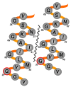
Figure 1
Figure 1
Schematic model of hypothetical glycine zipper-mediated interaction between Aβα-helical regions. Model of possible packing arrangement between C-terminal regions (residues 24-39) of neighboring Aβ molecules. The glycine zipper interface is represented by the jagged line; residue G37 is highlighted in red.
Figure 2
Figure 2
Reduced toxicity of Aβ G37L expressed in C. elegans. A. Representative plot of paralysis onset in transgenic worms induced to express Aβ wild type (strain CL4176) or Aβ G37L (strain CL3523). In both of these strains the transgenes are marked with the dominant Roller mutation rol-6(su1006). B. Paralysis plot for an independent pair of transgenic strains induced to express Aβ wild type (CL2659) or Aβ G37L (CL2621). In both of these strains the transgenes are marked with the intestinal GFP marker Pmtl-2::GFP. C. Representative immunoblot of worm protein extracts (20 μg/lane) probed with anti-Aβ monoclonal antibody 6E10. Lane 1, 25 ng synthetic Aβ1-42; lane 2, CL4176 (Aβ wild type); lane 3, CL2659 (Aβ wild type); lane 4, CL3523 (Aβ G37L); lane 5, CL2621 (Aβ G37L). Note that although the overall patterns of oligomeric Aβ bands were similar in strains expressing Aβ wild type or Aβ G37L, differences did appear in the region of dimeric Aβ (arrow). D. Immunostaining with mAb 6E10 of transgenic worms expressing Aβ wild type (upper panel) or Aβ G37L (lower panel). Nuclei stained with DAPI (blue), 6E10 immunoreactivity in red. Size bar = 10 μm. E. Electron micrographs of sections of transgenic worms fixed by high pressure freezing. Left panel, section through body wall muscle of worm expressing Aβ wild type (CL4176). Note abnormal "light" mitochondria (arrows). Right panel, equivalent section through body wall of transgenic worm expressing Aβ G37L (CL3523). Note normal dark mitochondria (arrows). S = sarcomeres, N = nucleus, M = mitochondria. Size bar = 1 μm.
Figure 3
Figure 3
Relative paralysis rates of worms expressing wild type or variant Aβ. Representative plot of transgenic worms induced to express Aβ. Strains used were: CL2654 (Aβ L17P), CL2659 (Aβ wild type), CL2666 (Aβ G29L G33L G37L), CL2621 (Aβ G37L) and CL2716 (Aβ G37F).
Figure 4
Figure 4
Effect of G37L substitution on Aβ mediated aggregation and β-amyloid formation. A. Digital overlay of differential interference contrast (DIC) and epifluorescence images of live transgenic worms expressing GFP::Aβ fusion proteins. Top panel, third larval stage worm expressing GFP::Aβwild type fusion protein (strain CL1332). Note GFP inclusions in body wall muscle. Middle panel, third larval stage worm expressing GFP::Aβ L17P fusion protein (strain CL1364). Note diffuse GFP in body wall muscle. Lower panel, third larval stage worm expressing GFP::AβG37L fusion protein. Note similar GFP inclusions as observed in worms expressing GFP::Aβ wild type (top panel). Size bar = 100 μm. B. Amyloid formation in transgenic worms with constitutive muscle expression of Aβwild type or G37L. DIC/epifluorescence images of live adult transgenic worms stained with amyloid-specific dye X-34 (33). Top panel, CL2006 (wild type Aβexpression). Note lateral amyloid deposits (small arrows) in body wall muscle and non-specific staining of the buccal cavity (large arrow). Lower panel, CL2564 (AβG37L). Muscle X-34 deposits are undetectable, although staining is still seen in the buccal cavity (large arrow). Size bar = 100 μm. C. Quantification of X-34 deposits in CL2006 (Aβwild type), CL2564 (AβG37L), and CL802 (control non-transgenic) worms. Error bars = S.E.M.
Figure 5
Figure 5
Toxicity of Aβ wild type and Aβ G37L peptides on Neuro 2a cells. Relative viability of Neuro 2a cells after treatment with Aβ wild type and Aβ G37L peptides for 24 hours, either singly or in various combinations. Viability was measured by the MTT assay and normalized to the vehicle-treated control cells. A. Neuro 2a cells were treated with 4 μM Aβ wild type that had been mixed with from 0-4 μM Aβ G37L for 20 min at RT before addition to the cells. The viability of each mixture was significantly different from that of Aβ wild type alone, and there was no significant difference in the viability of the vehicle control and an equimolar mixture of Aβ wild type and Aβ G37L (Student's t-test). Overall, Aβ G37L mitigated the toxicity of Aβ wild type in a linear dose dependent manner (R2 = 0.98). B. Neuro 2a cells were treated as described above with 4 μM Aβ wild type and/or 4 μM Aβ G37L. Note that the ability of Aβ G37L to inhibit Aβ wild type toxicity is lost when these peptides are oligomerized separately before addition to the Neuro 2a cell culture. (* P < 0.05, NS = not significant).
Figure 6
Figure 6
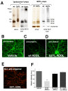
Toxicity of Aβ wild type and Aβ G37L oligomers (ADDLs) in primary hippocampal cultures. A. SDS-PAGE fractionation of oligomers formed from synthetic Aβ1-42 wild type or G37L. The left panel displays oligomers formed using the "globulomer" preparation of Barghorn et al [21]. The right panel displays oligomers formed using the "ADDL" preparation originally described by Lambert et al [24]. Monoclonal antibody 6E10 recognizes an epitope included in Aβresidues 16-24; mAb NU1 preferentially binds oligomeric Aβ. Note that for both oligomer preparations Aβ G37L tends to form higher molecular weight species. B - D. Representative epifluorescence images of anti-drebrin staining of embryonic rat hippocampal neurons treated after 21 days in culture for 24 hr with vehicle (B), 500 nM Aβ wild type ADDLs (C), or 500 nM Aβ G37L ADDLs (D). Note the strong reduction of drebrin staining is induced by exposure to wild type, but not G37L, ADDLs. E. Hippocampal neurons treated with Aβ G37 ADDLs, washed, fixed, and probed with anti-Aβ oligomer antibody NU1. Note robust binding of the Aβ G37L ADDLs to neurons. F. Quantification of drebrin immunoreactivity in hippocampal neurons exposed to Aβ wild type or G37L ADDLs. Exposure to Aβ wild type ADDLs significantly reduced drebrin immunoreactivity relative to vehicle-treated neurons (*P < 0.01), but no significant reduction was found for Aβ G37L ADDLs or ADDLs formed from a 1:1 mixture of Aβ wild type and G37L (Tukey Multiple Comparisons test).
Figure 7
Figure 7
Effect of Aβ wild type and G37L ADDL exposure on tau hyperphosphorylation. A. - D. Representative epifluorescence images of cultured embryonic hippocampal neurons that were exposed for 18 hr to vehicle (A), 500 nM Aβ G37L ADDLs (B), 500 nM Aβ scrambled ADDLs (C), or 500 nM Aβ wild type ADDLs (D), then fixed and probed with anti-phospho tau monoclonal antibody PHF-1. Note strong increase in PHF-1 staining only in neurons treated with Aβ wild type ADDLs. E. Quantification of PHF-1 staining. Exposure to Aβ wild type produced significant increases in PHF-1 staining relative to control at 4 hr (204% ± 13; p = 0.0023; n = 5) and 18 hr (416% ± 48; p = 0.0041; n = 5); none of the other treatments significantly increased PHF-1 staining (Student's T-test, * P < 0.005).
Figure 8
Figure 8
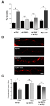
Toxicity of wild type and variant Aβ in adenovirus transfection model. Cultured rat cortical neurons were transfected with adenovirus engineered for doxycycline-inducible expression of wild type or variant Aβ, and toxicity was assayed by scoring of apoptotic nuclei or synaptophysin expression. A. Relative toxicity (pyknotic nuclei fraction) in neurons transfected with Aβ-expressing adenovirus with (+ Dox) and without induction. Transfection with adenovirus expressing Aβ wild type or Aβ L17P leads to significant toxicity after transgene induction (* P < 0.001), while transfection with adenovirus expressing Aβ G37L, or co-transfection with Aβ wild type and Aβ G37L, does not lead to significant toxicity after doxycycline induction. B. Representative processes containing synapses labeled with synaptophysin in Aβ wild type and Aβ G37L expressing neuronal cultures. Control panel refers to non-induced cultures. Scale bar, 5 μm. C. Analysis of puncta density (number of synapses per 25 μm of neurite) indicated fewer synapses in Aβ wild type expressing cells compared to non-induced controls. Intraneuronal Aβ G37L and co-expression of Aβ wild type and Aβ G37L resulted in no significant synapse alterations. Data from 3-5 different cultures from 3 independent isolations. n (neurites) = 154 Aβ wild type, 98 Aβ G37L, 118 Aβ wild type + G37L. (* = P < 0.001, NS = not significant, P > 0.1). Error bars represent SEM.
Figure 9
Figure 9
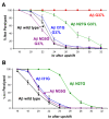
Second site mutations partially restore Aβ G37L toxicity. A. Paralysis plots of transgenic C. elegans expressing wild type, G37L, or double mutant Aβ. Note that the transgenic worms expressing the candidate compensatory mutations all paralyze faster than worms expressing Aβ G37L. B. Paralysis plot of transgenic C. elegans expressing single compensatory mutations. None of these single site mutations confer obvious hyper-toxicity.

Similar articles

Cited by

References

    1. Hardy J, Selkoe DJ. The amyloid hypothesis of Alzheimer's disease: progress and problems on the road to therapeutics. Science. 2002;297:353–356. doi: 10.1126/science.1072994. - DOI - PubMed
    1. Pappolla MA, Sos M, Omar RA, Bick RJ, Hickson-Bick DL, Reiter RJ, Efthimiopoulos S, Robakis NK. Melatonin prevents death of neuroblastoma cells exposed to the Alzheimer amyloid peptide. J Neurosci. 1997;17:1683–1690. - PMC - PubMed
    1. Hertel C, Terzi E, Hauser N, Jakob-Rotne R, Seelig J, Kemp JA. Inhibition of the electrostatic interaction between beta-amyloid peptide and membranes prevents beta-amyloid-induced toxicity. Proc Natl Acad Sci USA. 1997;94:9412–9416. doi: 10.1073/pnas.94.17.9412. - DOI - PMC - PubMed
    1. Yankner BA, Duffy LK, Kirschner DA. Neurotrophic and neurotoxic effects of amyloid beta protein: reversal by tachykinin neuropeptides. Science. 1990;250:279–282. doi: 10.1126/science.2218531. - DOI - PubMed
    1. Link CD. Expression of human beta-amyloid peptide in transgenic Caenorhabditis elegans. Proc Natl Acad Sci USA. 1995;92:9368–9372. doi: 10.1073/pnas.92.20.9368. - DOI - PMC - PubMed

LinkOut - more resources