. 2011 Aug 24:8:413.
doi: 10.1186/1743-422X-8-413.

Expression and characterization of UL16 gene from duck enteritis virus

Affiliations

Expression and characterization of UL16 gene from duck enteritis virus

Qin He et al. Virol J. .

Abstract

Background: Previous studies have indicated that the UL16 protein and its homologs from herpesvirus were conserved and played similar roles in viral DNA packaging, virion assembly, budding, and egress. However, there was no report on the UL16 gene product of duck enteritis virus (DEV). In this study, we analyzed the amino acid sequence of UL16 using bioinformatics tools and expressed in Escherichia coli Rosetta (DE3) induced by isopropy1-β-D-thiogalactopyranoside (IPTG). The recombinant protein was produced, purified using a Ni-NTA column and used to generate the polyclonal antibody against UL16. The intracellular distribution of the DEV UL16 product was carried out using indirect immunofluorescence assay.

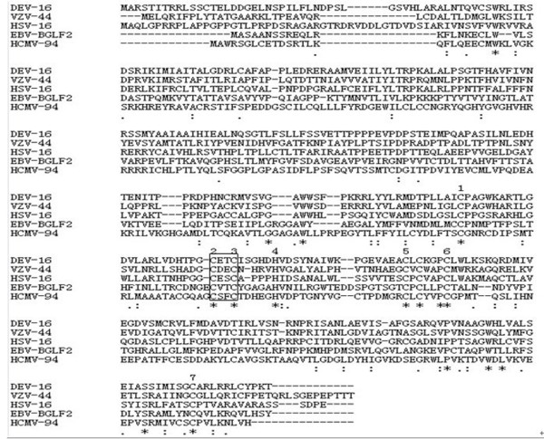
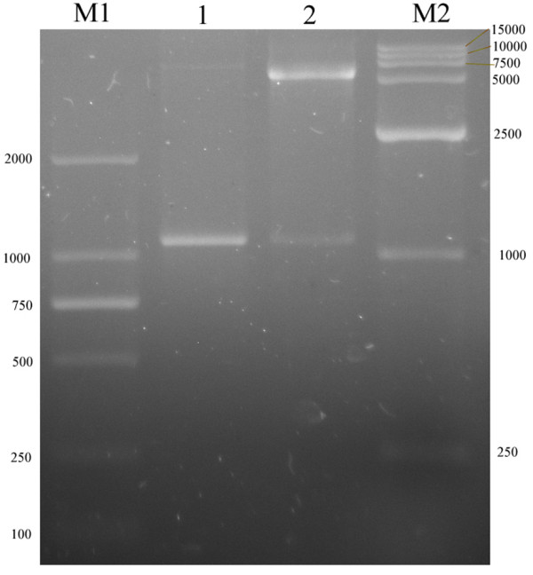
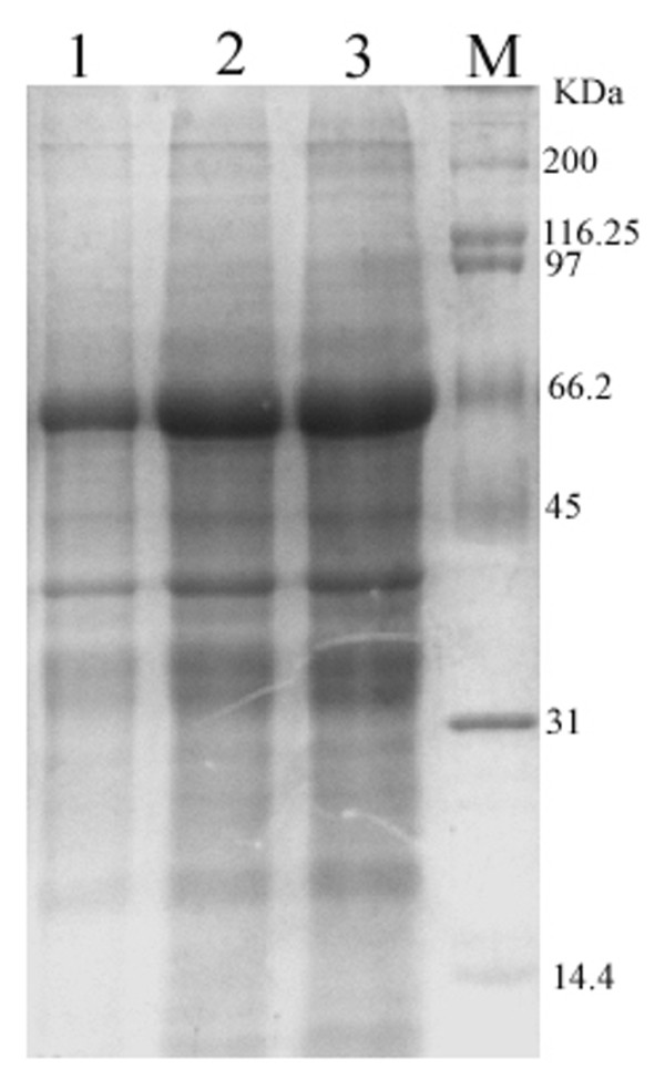
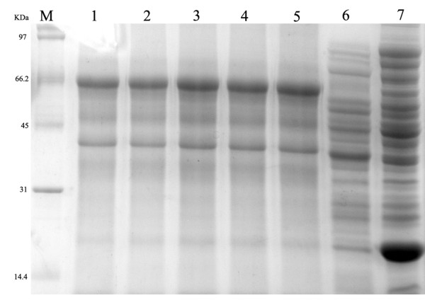
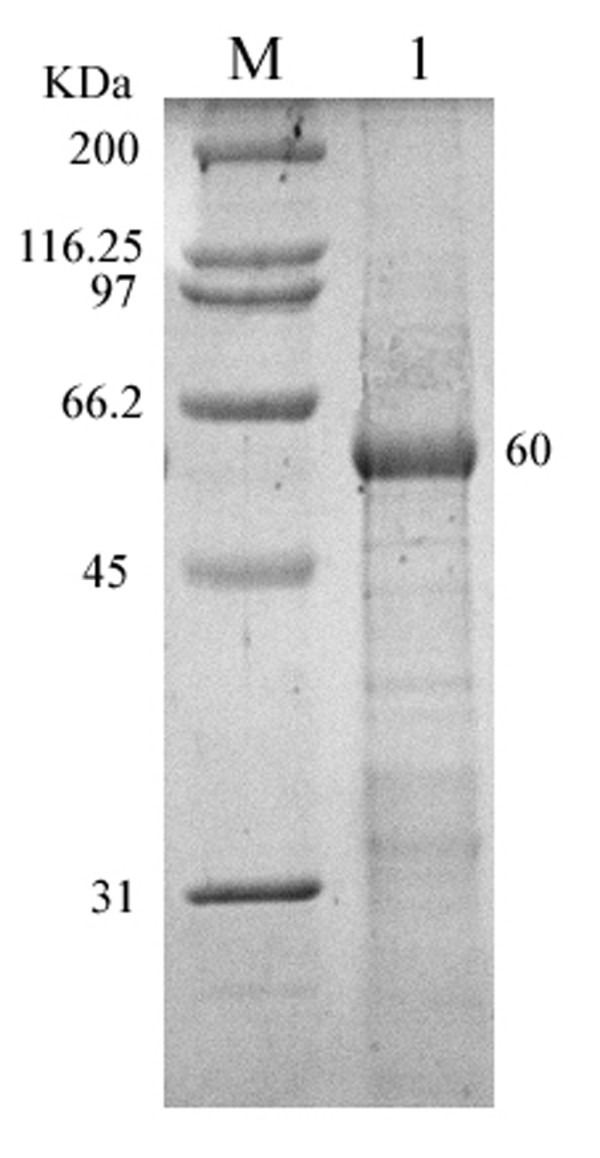
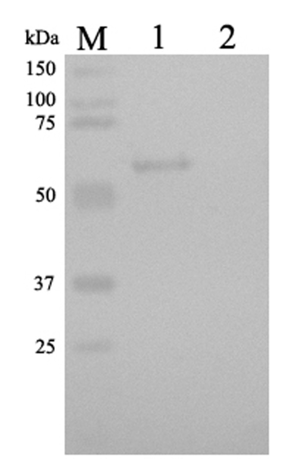
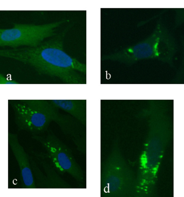
Results: In our study, UL16 gene of DEV was composed of 1089 nucleotides, which encoded 362 amino acids. Multiple sequence alignment suggested that the UL16 gene was highly conserved in herpesvirus family. The UL16 gene was cloned into a pET prokaryotic expression vector and transformed into Escherichia coli Rossetta (DE3) induced by IPTG. A 60kDa fusion protein band corresponding to the predicted size was produced on the SDS-PAGE, purified using a Ni-NTA column. Anti-UL16 polyclonal sera was prepared by immunizing rabbits, and reacted with a band in the IPTG induced cell lysates with an apparent molecular mass of 60 kDa. In vivo expression of the UL16 protein in DEV infected duck embryo fibroblast cells (DEFs) was localized mostly around perinuclear cytoplasmic area and in cytosol using indirect immunofluorescence assay.

Conclusions: The UL16 gene of DEV was successfully cloned, expressed and detected in DEV infected DEFs for the first time. The UL16 protein localized mostly around perinuclear cytoplasmic area and in cytosol in DEV infected DEFs. DEV UL16 shared high similarity with UL16 family members, indicating that DEV UL16 many has similar function with its homologs. All these results may provide some insight for further research about full characterizations and functions of the DEV UL16.


Figures

Figure 1
Figure 1
Amino acid sequence comparison between the putative proteins encoded by DEV UL16 and it homologs: HSV-1 UL16, VZV ORF44, EBV BGLF2, and HCMV UL94. Sequences were aligned with the Clustalx1.8 software. Absence of amino acid is shown by dash '-' in the sequences while '*', ':', and '.' indicate identical amino acid residues, conserved residues and semi-conserved residues in all sequence used in the alignment respectively.
Figure 2
Figure 2
Identification of recombination vector pET 32b (+)-UL16 by restriction enzymes digestion. Lane M1, DNA marker; Lane 1, PCR products from pET 32b(+)-UL16; Lane 2, recombination plasmid pET 32b(+)-UL16 was digested with two restriction enzymes HindIII and XhoI; Lane M2, DNA marker.
Figure 3
Figure 3
SDS-PAGE analysis of the expression results of recombinant plasmid pET32b-UL16 in different temperature from Rossetta(DE3). Lane M = Protein marker; lanes 1-4 = cells were grown respectively at 25, 30, 37°C after induction with IPTG.
Figure 4
Figure 4
SDS-PAGE analysis of different time course of recombinant plasmid pET32b-UL16 production from Rossetta(DE3). Lane M = Protein marker; lanes 1 = the plasmid pET32b (+) was induced with IPTG; Lane 4 = the recombinant plasmid pET32b-UL16 was uninduced; Lane 2, 3, 5, 6 = cells were grown respectively for 6, 8, 4 and 2 h after induction with IPTG.
Figure 5
Figure 5
Production of recombinant plasmid pET32b-UL16 from Rossetta(DE3) in different IPTG concentrations. Lanes 1-6 = cells were grown and induced at 1.0, 0.8, 0.6, 0.4, 0.2 and 0.0 mmol/l IPTG, Lane 7 = the plasmid pET32b (+) was induced with IPTG.
Figure 6
Figure 6
SDS-PAGE analysis of the purity of recombinant plasmid pET32b-UL16 from Rossetta (DE3). Lane M = Protein marker; lanes 1 = recombinant protein from Rossetta (DE3) purified by the Ni+-NTA agarose gel.
Figure 7
Figure 7
Western blotting identification of recombinant protein UL16 expressed in Escherichia coli Rossetta (DE3) with the rabbit serum of anti-DEV UL16 IgG. lane 1 = expression of recombinant plasmid pET32b-UL16 induced by IPTG; Lane 2 = Expression of recombinant plasmid pET32b-UL16 uninduced Escherichia coli Rossetta (DE3) cell lysate.
Figure 8
Figure 8
Intracellular localization of the UL16 protein in DEV CHv infected DEFs. Mock- and DEV CHv infected cells were fixed at the indicated times postinfection and processed for indirect immunofluorescence. Mock-infected cells were fixed after 36 h incubation (a); DEV CHv infected cells were fixed at 18(b), 24(c), and 36 h postinfection (d). ×400.

References

    1. Davison S, Converse KA, Hamir AN, Eckroade RJ. Duck viral enteritis in domestic Muscovy ducks in Pennsylvania. Avian Dis. 1993;37:1142–1146. doi: 10.2307/1591927. - DOI - PubMed
    1. Campagnolo ER, Baneriee M, Paniqrahy B, Jones RL. An outbreak of duck viral enteritis (duck plague) in domestic Muscovy ducks (Cairina moschata domesticus) in Illinois. Avian Dis. 2001;45:522–528. doi: 10.2307/1592999. - DOI - PubMed
    1. Fadly AM, Glisson JR, McDougald LR, Nolan Lk, Swayne DE. Diseases of Poultry American. Wiley-BlackwellSaif YM; 2008. Duck Virus Enteritis; pp. 384–393.
    1. Baudet AE. Mortality in ducks in the Netherlands caused by a filtrable virus; fowl plague. Tijdschr Diergeneeskd. 1923;50:455–459.
    1. Leibovitz L, Hwang J. Duck plague on the American continent. Avian Dis. 1968;12:361–378. doi: 10.2307/1588237. - DOI - PubMed

Publication types

MeSH terms

LinkOut - more resources