

Effects of Solanum glaucophyllum toxicity on cell proliferation and apoptosis in the small and large intestine of rabbits
- PMID: 21862088
- PMCID: PMC7125838
- DOI: 10.1016/j.rvsc.2011.07.018
Effects of Solanum glaucophyllum toxicity on cell proliferation and apoptosis in the small and large intestine of rabbits
Abstract
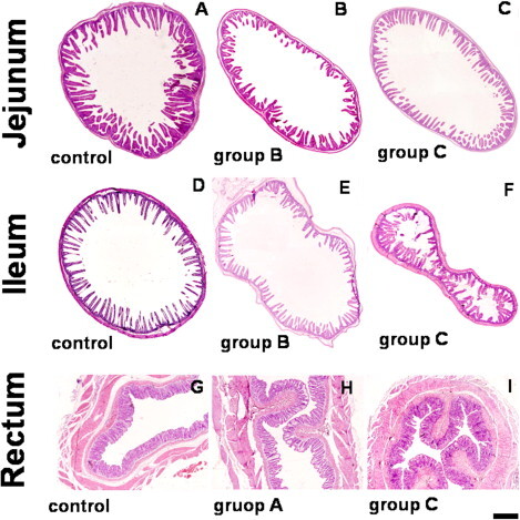
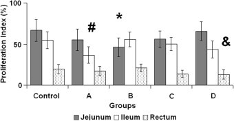
Vitamin D regulates mineral homeostases and enterocyte proliferation and differentiation. Hypervitaminosis D generates changes in cell proliferation, differentiation and apoptosis in several organs. We analysed morphometric parameters and proliferative and apoptotic indices in the intestinal epithelium of rabbits with hypervitaminosis D induced by the chronic treatment with the calcinogenic plant Solanum glaucophyllum. Rabbits were treated for 15 or 30 days. A group was treated for 15 days and led to possible recovery for 30 days. Another group was nutritionally restricted for 30 days. Morphological, morphometric, proliferative and apoptotic changes were found in the treated animals. Mild atrophy and reduced proliferation was found in the jejunum and ileum. Apoptosis increased in the crypts of the ileum and in the superficial epithelium and crypts of the rectum. Most of the alterations were partially recovered. The possible involvement in these changes of the hypervitaminosis D-like state induced by S. glaucophyllum is discussed.
Copyright © 2011 Elsevier Ltd. All rights reserved.
Figures


Similar articles
-
Glycoconjugate histochemistry in the small and large intestine of normal and Solanum glaucophyllum-intoxicated rabbits.Res Vet Sci. 2010 Oct;89(2):214-22. doi: 10.1016/j.rvsc.2010.03.002. Epub 2010 Mar 29. Res Vet Sci. 2010. PMID: 20350732
-
Paneth cells: histochemical and morphometric study in control and Solanum glaucophyllum intoxicated rabbits.Eur J Histochem. 2008 Apr-Jun;52(2):93-100. doi: 10.4081/1193. Eur J Histochem. 2008. PMID: 18591155
-
Effect of the addition of different concentrations of Solanum glaucophyllum desf. extract on chondrocyte cultures from the growth cartilage of newborn rats.Toxicon. 2023 Jul;230:107158. doi: 10.1016/j.toxicon.2023.107158. Epub 2023 May 11. Toxicon. 2023. PMID: 37172829
-
Peptide YY induces intestinal proliferation in peptide YY knockout mice with total enteral nutrition after massive small bowel resection.J Pediatr Gastroenterol Nutr. 2009 May;48(5):517-25. doi: 10.1097/MPG.0b013e31818c5fd8. J Pediatr Gastroenterol Nutr. 2009. PMID: 19367178
-
The effect of leptin on intestinal recovery following ischemia-reperfusion injury in a rat.Pediatr Surg Int. 2007 May;23(5):473-8. doi: 10.1007/s00383-006-1863-9. Pediatr Surg Int. 2007. PMID: 17203324
Cited by
-
Use of vitamin d3 and its metabolites in broiler chicken feed on performance, bone parameters and meat quality.Asian-Australas J Anim Sci. 2013 Mar;26(3):408-15. doi: 10.5713/ajas.2012.12455. Asian-Australas J Anim Sci. 2013. PMID: 25049804 Free PMC article.
-
Targeted delivery of 1,25-dihydroxyvitamin D3 to colon tissue and identification of a major 1,25-dihydroxyvitamin D3 glycoside from Solanumglaucophyllum plant leaves.J Steroid Biochem Mol Biol. 2015 Apr;148:318-25. doi: 10.1016/j.jsbmb.2014.10.019. Epub 2014 Nov 1. J Steroid Biochem Mol Biol. 2015. PMID: 25445916 Free PMC article. Review.
-
The Genus Solanum: An Ethnopharmacological, Phytochemical and Biological Properties Review.Nat Prod Bioprospect. 2019 Apr;9(2):77-137. doi: 10.1007/s13659-019-0201-6. Epub 2019 Mar 12. Nat Prod Bioprospect. 2019. PMID: 30868423 Free PMC article. Review.
-
Enzootic calcinosis in horses grazing Solanum glaucophyllum in Argentina.J Vet Diagn Invest. 2018 Mar;30(2):286-289. doi: 10.1177/1040638717746447. Epub 2017 Dec 4. J Vet Diagn Invest. 2018. PMID: 29202673 Free PMC article.
-
The effects of in ovo injected vitamin D3 sources on the eggshell temperature and early posthatch performance of Ross 708 broilers,.Poult Sci. 2020 Mar;99(3):1357-1362. doi: 10.1016/j.psj.2019.10.055. Epub 2019 Dec 12. Poult Sci. 2020. PMID: 32115025 Free PMC article.
References
-
- Bancroft J.D., Stevens A. Third ed. Churchill; Livingstone, New York: 1990. Theory and Practice of Histological Techniques. pp. 639–640.
-
- Barros S.S., Gimeno E.J. Cell differentiation and bone protein synthesis in the lungs of sheep with spontaneous calcinosis. Journal of Comparative Pathology. 2000;123:270–277. - PubMed
-
- Bikle D.D. What is new in vitamin D: 2006–2007. Current Opinion in Rheumatology. 2007;19:383–388. - PubMed
-
- Biol-N’ Garagba M.C., Greco S., George P., Hugueny I., Louisot P. Polyamine participation in the maturation of glycoprotein fucosylation, but not sialylation, in rat small intestine. Pediatric Research. 2002;51:625–634. - PubMed
Publication types
MeSH terms
Substances
LinkOut - more resources
Full Text Sources
