. 2011 Sep;138(18):3921-30.
doi: 10.1242/dev.062554.

Lef1 is required for progenitor cell identity in the zebrafish lateral line primordium

Affiliations

Lef1 is required for progenitor cell identity in the zebrafish lateral line primordium

Hillary F McGraw et al. Development. 2011 Sep.

Abstract

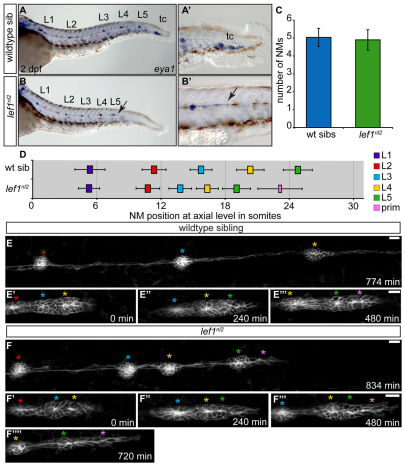
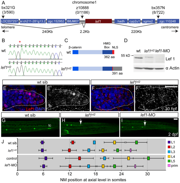
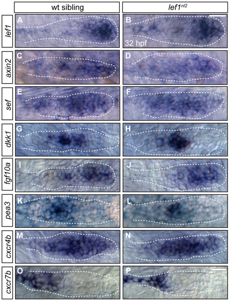
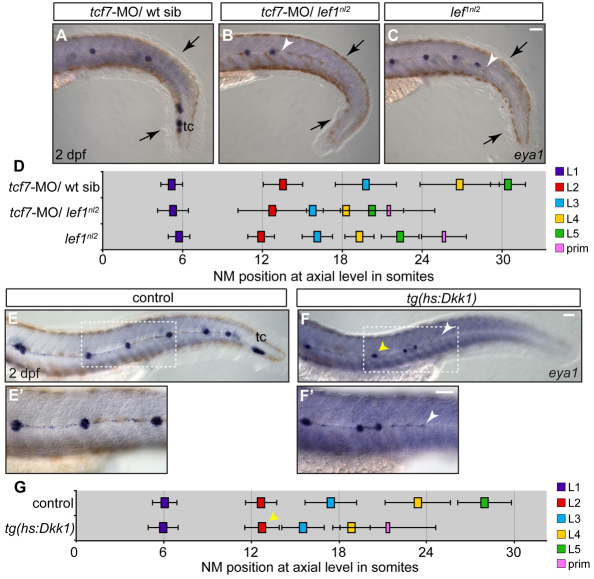
The zebrafish posterior lateral line (pLL) is a sensory system that comprises clusters of mechanosensory organs called neuromasts (NMs) that are stereotypically positioned along the surface of the trunk. The NMs are deposited by a migrating pLL primordium, which is organized into polarized rosettes (proto-NMs). During migration, mature proto-NMs are deposited from the trailing part of the primordium, while progenitor cells in the leading part give rise to new proto-NMs. Wnt signaling is active in the leading zone of the primordium and global Wnt inactivation leads to dramatic disorganization of the primordium and a loss of proto-NM formation. However, the exact cellular events that are regulated by the Wnt pathway are not known. We identified a mutant strain, lef1(nl2), that contains a lesion in the Wnt effector gene lef1. lef1(nl2) mutants lack posterior NMs and live imaging reveals that rosette renewal fails during later stages of migration. Surprisingly, the overall primordium patterning, as assayed by the expression of various markers, appears unaltered in lef1(nl2) mutants. Lineage tracing and mosaic analyses revealed that the leading cells (presumptive progenitors) move out of the primordium and are incorporated into NMs; this results in a decrease in the number of proliferating progenitor cells and eventual primordium disorganization. We concluded that Lef1 function is not required for initial primordium organization or migration, but is necessary for proto-NM renewal during later stages of pLL formation. These findings revealed a novel role for the Wnt signaling pathway during mechanosensory organ formation in zebrafish.


Figures

Fig. 1.
Fig. 1.
Abnormal primordium patterning leads to loss of terminal neuromasts in the lef1nl2 mutant. (A,B) eya1 expression in the pLL of wild-type sibling and lef1nl2 mutant embryos at 2 dpf. The lef1nl2 mutant lacks a tc. (A′,B′) Distal limit of eya1 expression in wild-type and lef1nl2 mutant (arrow) embryos. (C) Average number of NMs (excluding tc) in lef1nl2 mutant and wild-type sibs (mean±s.d.) are not significantly different at 2 dpf (n=28 wild type, 11 lef1nl2, P=0.49, Student's t-test). (D) Axial positions of L1-L5 in lef1nl2 and wild-type siblings at 2 dpf (mean±s.d.). The axial level of stalled primordium (prim) in lef1nl2 mutants is indicated by the pink bar. Position of L1-L5 is significantly shifted anteriorly in lef1nl2 mutants (n=18, P<0.001, two-way ANOVA with replication). (E-F′′) Stills from time-lapse movies of primordium migration in wild-type sibling and lef1nl2 mutant embryos that express the Tg(–8.0cldnb:lynGFP) transgene. Wild-type and lef1nl2 embryos were imaged beginning at 34 hpf for 780 minutes and 840 minutes, respectively (see Movies 1 and 2 in the supplementary material). (E-E′′) Over the course of 780 minutes, the primordium in the wild-type embryo migrated out of frame, deposited three NMs (red, blue and yellow asterisks) and generated two new rosettes (green and pink asterisks). (F-F′′′) Over the course of 840 minutes, the primordium in the lef1nl2 mutant has slowed and became elongated (pink asterisk), having deposited four NMs (red, blue, yellow and green asterisks). Scale bars: 20 μm.
Fig. 2.
Fig. 2.
The lef1nl2 mutant contains a lesion in the lef1 gene. (A) The lef1nl2 mutation was mapped to a 460 Kb region on chromosome 1. Numbers of recombinants are indicated under each marker. (B) The lef1nl2 mutation is a single guanine insertion at position 1120 (red asterisk). (C) The resulting frame shift is predicted to disrupt the nuclear localization signal (NLS) and extend the protein by 29 amino acids (aa; also shown a β-catenin binding domain and HMG Box). (D) Western blot of wild-type, lef1nl2 mutant and lef1 morphant whole embryo lysates probed with anti-Lef1 antibody and anti-α actin antibody. (E-F′) Immunolabeling using anti-Lef1 antibody revealed nuclear labeling (red) in wild-type sibling and cytoplasmic labeling in lef1nl2 mutant embryos. Nuclei are labeled with DAPI. (G-I) Wild-type sibling, lef1nl2 mutant and lef1-MO injected embryos expressing the Tg(–8.0cldnb:lynGFP) transgene at 2 dpf. (G) The pLL was truncated prematurely (white arrows) in the lef1nl2 mutant (H) and lef1 morphant (I). (J) The axial positions of the deposited NMs are shifted anteriorally in lef1nl2 mutants when compared with wild-type siblings and in lef1 morphants when compared with uninjected controls. (Data presented as mean±s.d.; n=6-11 P<0.001, two-way ANOVA with replication.) Scale bars: 50 μm.
Fig. 3.
Fig. 3.
Primordium patterning is maintained in lef1nl2 mutants. (A-P) RNA in situ hybridization of factors required for primordium patterning in wild-type siblings (left panels) and lef1nl2 mutants (right panels) at 32 hpf. Expression of lef1 (A,B), axin2 (C,D), sef (E,F), dkk1 (G,H), fgf10a (I,J), pea3 (K,L), cxcr4b (M,N) and cxcr7b (O,P) are similar in both wild-type and mutant embryos. Scale bars: 20 μm.
Fig. 4.
Fig. 4.
Combined loss of Lef1 and Tcf7 does not recapitulate the pLL phenotype seen with the complete loss of Wnt signaling. (A-C) Zygotes from a lef1nl2/+ incrosses were injected with tcf7-MO. NM distribution in the injected embryos was assessed by eya1 expression at 2 dpf and compared with uninjected lef1 mutant embryos. The loss of Tcf7 alone did not affect pLL patterning in lef1nl2/+ sibling (A), the pLL is truncated more severely lef1nl2/–; tcf7-MO (B; arrowhead) compared with lef1nl2 mutants alone (C; arrowhead). Note reduced fin fold (black arrows) caused by a combined loss of Lef1 and Tcf7 as reported previously (Nagayoshi et al., 2008). (D) Axial positions of the pLL NMs (L1-L5) and primordium (mean±s.d.; n=11, P<0.001, two-way ANOVA with replication). (E-F′) pLL formation in 2 dpf embryos following the induction of Dkk1 in the Tg(hsp70l:dkk1-GFP) line by a heat-shock at 28 hpf. Position of the primordium at the time of heat-shock is marked by the yellow arrowheads. Dashed rectangles indicate the regions shown at higher magnification in E′,F′. Following the heat-shock, one or two NMs are deposited and the pLL ends with a trail of eya1-positive cells (white arrowheads). (G) Axial positions of NMs deposited following heat-shock in control and Tg(hsp70l:dkk1-GFP) embryos (n=11). Scale bars: 20 μm.
Fig. 5.
Fig. 5.
Lef1 function is required in leading edge cells of the primordium for proper pLL formation. (A-C′) Confocal projection obtained from a lef1nl2 mutant host that received wild-type donor cells (rhodamine dextran, red). (A) At 24 hpf, the primordium contains donor cells in the leading zone and caudal-most rosette. (B) The same embryo as in A, showing complete primordium migration and tc formation at 48 hpf. (B′) High magnification of region outlined in B; the primordium is entirely composed of donor cells. (C) Lateral line is truncated on the contralateral side of the same embryo. (C′) High magnification of the region outlined in C. Both wild-type donors and mutant hosts expressed Tg(–8.0cldnb:lynGFP) transgene. Scale bars: 20 μm.
Fig. 6.
Fig. 6.
Wnt signaling regulates proliferation in the primordium. (A-C′′) Confocal projections of primordia following BrdU incorporation between 32.5 and 34.0 hpf in wild-type, lef1nl2 mutant and Tg(hsp70l:dkk1-GFP) embryos. All embryos express Tg(–8.0cldnb:lynGFP). BrdU incorporation in the primordia of the wild-type (A-A′′), the lef1nl2 mutant (B-B′′) and the Tg(hsp70l:dkk1-GFP) transgenic embryos heat-shocked at 28 hpf (C-C′′). Scale bar: 20 μm. (D,E) BrdU incorporation index for wild-type sibling and lef1nl2 mutants (D) and control and Tg(hsp70l:dkk1-GFP) embryos (E). (F,G) BrdU incorporation index for leading region in wild-type siblings and lef1nl2 mutants (F) and controls and Tg(hsp70l:dkk1-GFP) embryos (G) (n=10-22 embryos; data presented as mean±s.e.m., **P<0.009, ***P<0.002, Student's t-test). The leading region is defined as the cells posterior to the leading rosette in the primordium.
Fig. 7.
Fig. 7.
Leading region cells change their fate in the absence of Lef1 function. (A-C′) Tg(–8.0cldnb:lynGFP) zygotes were injected with a nuclear-localized Kaede mRNA and an average of two cells were photoconverted at 24 hpf. (A,B) Wild-type and lef1nl2 mutant embryos immediately following photoconversion are shown. (C-D′) The embryos in A-B′ at 48 hpf showing the location of the progeny of the photoconverted cells. Note absence of the labeled (red) cells in the leading region of lef1nl2 mutant embryos. (E) Quantification of converted cells at 24 hpf and their progeny at 48 hpf. (F) Localization of converted cells at 48 hpf in wild-type and lef1nl2 mutants. Cells in lef1nl2 mutants are significantly more likely to be localized in NMs and not in the primordia when compared with wild type (mean±s.e.m., n=27 wild-type and 16 lef1nl2 mutant embryos, *P<0.04, Student's t-test). (G-H′′) Still images from time-lapse movies (see Movies 3 and 4 in the supplementary material) demonstrating division of Kaede-positive cells (red) in a wild-type (G-G′′) and a lef1nl2 embryos (H-H′′). Specific time points were chosen to show a subset of labeled cells just before and after cell divisions. Leading zone divisions marked by blue arrows, whereas cell divisions in rosettes are marked by yellow arrowheads. Scale bars: 20 μm.
Fig. 8.
Fig. 8.
Lef1 is required to maintain progenitor cell identity. (A-D) Confocal projections of Tg(–8.0cldnb:lynGFP)-positive, mosaic embryos containing either wild-type or lef1nl2 donor cells (red) at 24 and 48 hpf. (A) Wild-type host primordium at 24 hpf with wild-type donor cells in the leading region (yellow bracket). (B) Chimeric primordium containing lef1nl2 mutant cells in the leading region at 24 hpf (yellow bracket). At 48 hpf, wild-type donor cells remain in the leading region (C; yellow bracket), whereas lef1nl2 cells have moved out of the leading region (D; yellow bracket). (E-F′) Confocal projections of chimeric primordia containing wild type Tg(hsp70l:dkk1-GFP) donor cells. Embryos were heat-shocked at 28 hpf. (E) Primordium containing wild-type cells and a characteristic rounded morphology. (F) Wild-type primordium containing Tg(hsp70l:dkk1-GFP) donor cells shows loss of primordium organization. (F′) Contralateral side of the chimera shown in F is normal. Scale bars: 20 μm.

Similar articles

Cited by

References

    1. Abramoff M. D., Magelhaes P. J., Ram S. J. (2004). Image processing with ImageJ. Biophotonics Int. 11, 36-42
    1. Aman A., Piotrowski T. (2008). Wnt/beta-catenin and Fgf signaling control collective cell migration by restricting chemokine receptor expression. Dev. Cell 15, 749-761 - PubMed
    1. Aman A., Piotrowski T. (2009). Multiple signaling interactions coordinate collective cell migration of the posterior lateral line primordium. Cell Adh. Migr. 3, 365-368 - PMC - PubMed
    1. Aman A., Nguyen M., Piotrowski T. (2010). Wnt/β-catenin dependent cell proliferation underlies segmented lateral line morphogenesis. Dev. Biol. 349, 470-482 - PubMed
    1. Andermann P., Ungos J., Raible D. W. (2002). Neurogenin1 defines zebrafish cranial sensory ganglia precursors. Dev. Biol. 251, 45-58 - PubMed

Publication types

MeSH terms